Mohd Nurulhuda, Razali Masfueh, Ghazali Mariyam Jameelah, Abu Kasim Noor Hayaty
Department of Restorative Dentistry, Faculty of Dentistry, Universiti Kebangsaan Malaysia, Jalan Raja Muda Abdul Aziz, Kuala Lumpur 50300, Malaysia.
Department of Mechanical & Manufacturing Engineering, Faculty of Engineering & Built Environment, Universiti Kebangsaan Malaysia, Bangi 43600, Selangor, Malaysia.
Materials (Basel). 2022 Apr 2;15(7):2621. doi: 10.3390/ma15072621.
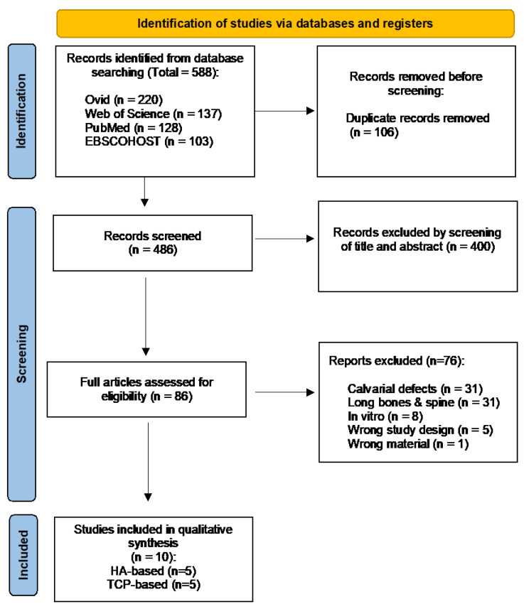
Three-dimensional-printed scaffolds have received greater attention as an attractive option compared to the conventional bone grafts for regeneration of alveolar bone defects. Hydroxyapatite and tricalcium phosphates have been used as biomaterials in the fabrication of 3D-printed scaffolds. This scoping review aimed to evaluate the potential of 3D-printed HA and calcium phosphates-based scaffolds on alveolar bone regeneration in animal models. The systematic search was conducted across four electronic databases: Ovid, Web of Science, PubMed and EBSCOHOST, based on PRISMA-ScR guidelines until November 2021. The inclusion criteria were: (i) animal models undergoing alveolar bone regenerative surgery, (ii) the intervention to regenerate or augment bone using 3D-printed hydroxyapatite or other calcium phosphate scaffolds and (iii) histological and microcomputed tomographic analyses of new bone formation and biological properties of 3D-printed hydroxyapatite or calcium phosphates. A total of ten studies were included in the review. All the studies showed promising results on new bone formation without any inflammatory reactions, regardless of the animal species. In conclusion, hydroxyapatite and tricalcium phosphates are feasible materials for 3D-printed scaffolds for alveolar bone regeneration and demonstrated bone regenerative potential in the oral cavity. However, further research is warranted to determine the scaffold material which mimics the gold standard of care for bone regeneration in the load-bearing areas, including the masticatory load of the oral cavity.
与传统骨移植相比,三维打印支架作为牙槽骨缺损再生的一种有吸引力的选择受到了更多关注。羟基磷灰石和磷酸三钙已被用作制造三维打印支架的生物材料。本综述旨在评估三维打印的基于羟基磷灰石和磷酸钙的支架在动物模型中牙槽骨再生的潜力。根据PRISMA-ScR指南,在四个电子数据库(Ovid、Web of Science、PubMed和EBSCOHOST)中进行了系统检索,截至2021年11月。纳入标准为:(i)接受牙槽骨再生手术的动物模型;(ii)使用三维打印的羟基磷灰石或其他磷酸钙支架进行骨再生或增强的干预措施;(iii)对三维打印的羟基磷灰石或磷酸钙的新骨形成和生物学特性进行组织学和微计算机断层扫描分析。本综述共纳入十项研究。所有研究均显示,无论动物种类如何,在新骨形成方面都取得了有前景的结果,且无任何炎症反应。总之,羟基磷灰石和磷酸三钙是用于三维打印牙槽骨再生支架的可行材料,并在口腔中显示出骨再生潜力。然而,有必要进一步研究以确定在包括口腔咀嚼负荷在内的承重区域中模拟骨再生护理金标准所需的支架材料。