• 文献检索
  • 文档翻译
  • 深度研究
  • 学术资讯
  • Suppr Zotero 插件Zotero 插件
  • 邀请有礼
  • 套餐&价格
  • 历史记录
应用&插件
Suppr Zotero 插件Zotero 插件浏览器插件Mac 客户端Windows 客户端微信小程序
定价
高级版会员购买积分包购买API积分包
服务
文献检索文档翻译深度研究API 文档MCP 服务
关于我们
关于 Suppr公司介绍联系我们用户协议隐私条款
关注我们

Suppr 超能文献

核心技术专利:CN118964589B侵权必究
粤ICP备2023148730 号-1Suppr @ 2026

文献检索

告别复杂PubMed语法,用中文像聊天一样搜索,搜遍4000万医学文献。AI智能推荐,让科研检索更轻松。

立即免费搜索

文件翻译

保留排版,准确专业,支持PDF/Word/PPT等文件格式,支持 12+语言互译。

免费翻译文档

深度研究

AI帮你快速写综述,25分钟生成高质量综述,智能提取关键信息,辅助科研写作。

立即免费体验

基于 OCTA 的眼前节 OCT 容积血流成像中空间-时间斑点方差对比

Spatial-Temporal Speckle Variance in the En-Face View as a Contrast for Optical Coherence Tomography Angiography (OCTA).

机构信息

Department of Internal Medicine, University of Texas Medical Branch at Galveston, 301 University Blvd, Galveston, TX 77555, USA.

Department of Ophthalmology and Visual Sciences, University of Texas Medical Branch at Galveston, 301 University Blvd, Galveston, TX 77555, USA.

出版信息

Sensors (Basel). 2022 Mar 22;22(7):2447. doi: 10.3390/s22072447.

DOI:10.3390/s22072447
PMID:35408061
原文链接:https://pmc.ncbi.nlm.nih.gov/articles/PMC9003003/
Abstract

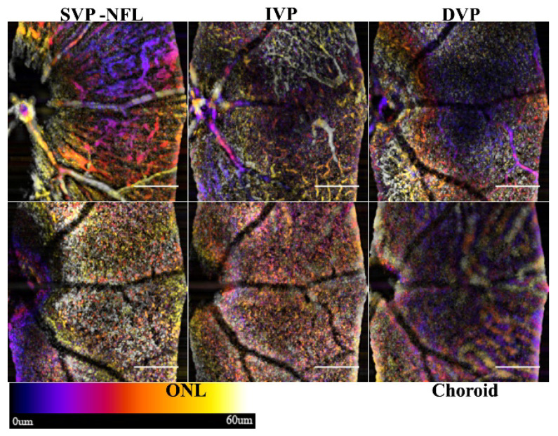
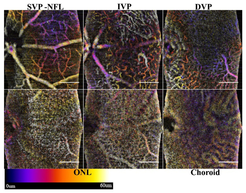
Optical Coherence Tomography (OCT) is an adaptable depth-resolved imaging modality capable of creating a non-invasive 'digital biopsy' of the eye. One of the latest advances in OCT is optical coherence tomography angiography (OCTA), which uses the speckle variance or phase change in the signal to differentiate static tissue from blood flow. Unlike fluorescein angiography (FA), OCTA is contrast free and depth resolved. By combining high-density scan patterns and image processing algorithms, both morphometric and functional data can be extracted into a depth-resolved vascular map of the retina. The algorithm that we explored takes advantage of the temporal-spatial relationship of the speckle variance to improve the contrast of the vessels in the en-face OCT with a single frame. It also does not require the computationally inefficient decorrelation of multiple A-scans to detect vasculature, as used in conventional OCTA analysis. Furthermore, the spatial temporal OCTA (ST-OCTA) methodology tested offers the potential for post hoc analysis to improve the depth-resolved contrast of specific ocular structures, such as blood vessels, with the capability of using only a single frame for efficient screening of large sample volumes, and additional enhancement by processing with choice of frame averaging methods. Applications of this method in pre-clinical studies suggest that the OCTA algorithm and spatial temporal methodology reported here can be employed to investigate microvascularization and blood flow in the retina, and possibly other compartments of the eye.

摘要

光学相干断层扫描(OCT)是一种适应性强的深度分辨成像方式,能够对眼睛进行非侵入性的“数字活组织检查”。OCT 的最新进展之一是光相干断层扫描血管造影术(OCTA),它利用信号中的散斑方差或相位变化来区分静态组织和血流。与荧光素血管造影(FA)不同,OCTA 是无对比且深度分辨的。通过结合高密度扫描模式和图像处理算法,可以将形态和功能数据提取为视网膜的深度分辨血管图。我们探索的算法利用了散斑方差的时空关系,以提高单次帧中 en-face OCT 中血管的对比度。它也不需要像传统的 OCTA 分析那样,通过多个 A 扫描的去相关来检测血管,这是计算效率低下的。此外,所测试的时空 OCTA(ST-OCTA)方法提供了事后分析的潜力,以提高特定眼部结构(如血管)的深度分辨对比度,其仅使用单个帧即可对大样本量进行高效筛选,并通过选择帧平均方法进行处理来进一步增强。该方法在临床前研究中的应用表明,本文报道的 OCTA 算法和时空方法可用于研究视网膜中的微血管化和血流,以及可能的眼部其他部位。

https://cdn.ncbi.nlm.nih.gov/pmc/blobs/e185/9003003/d43abb6a9db7/sensors-22-02447-g015.jpg
https://cdn.ncbi.nlm.nih.gov/pmc/blobs/e185/9003003/cb9de63293a0/sensors-22-02447-g001.jpg
https://cdn.ncbi.nlm.nih.gov/pmc/blobs/e185/9003003/2ed623099edf/sensors-22-02447-g002.jpg
https://cdn.ncbi.nlm.nih.gov/pmc/blobs/e185/9003003/f8556a962988/sensors-22-02447-g003.jpg
https://cdn.ncbi.nlm.nih.gov/pmc/blobs/e185/9003003/21008500535a/sensors-22-02447-g004.jpg
https://cdn.ncbi.nlm.nih.gov/pmc/blobs/e185/9003003/c03f2177f923/sensors-22-02447-g005.jpg
https://cdn.ncbi.nlm.nih.gov/pmc/blobs/e185/9003003/991cc3e0ece6/sensors-22-02447-g006.jpg
https://cdn.ncbi.nlm.nih.gov/pmc/blobs/e185/9003003/019d6045abfb/sensors-22-02447-g007.jpg
https://cdn.ncbi.nlm.nih.gov/pmc/blobs/e185/9003003/eb490cd8ed22/sensors-22-02447-g008.jpg
https://cdn.ncbi.nlm.nih.gov/pmc/blobs/e185/9003003/ce0d0081968f/sensors-22-02447-g009.jpg
https://cdn.ncbi.nlm.nih.gov/pmc/blobs/e185/9003003/b592926e9765/sensors-22-02447-g010.jpg
https://cdn.ncbi.nlm.nih.gov/pmc/blobs/e185/9003003/d223a8f63452/sensors-22-02447-g011.jpg
https://cdn.ncbi.nlm.nih.gov/pmc/blobs/e185/9003003/43c5e3a85446/sensors-22-02447-g012.jpg
https://cdn.ncbi.nlm.nih.gov/pmc/blobs/e185/9003003/dd6c9ced603e/sensors-22-02447-g013.jpg
https://cdn.ncbi.nlm.nih.gov/pmc/blobs/e185/9003003/1b5d35deb4ee/sensors-22-02447-g014.jpg
https://cdn.ncbi.nlm.nih.gov/pmc/blobs/e185/9003003/d43abb6a9db7/sensors-22-02447-g015.jpg

相似文献

1
Spatial-Temporal Speckle Variance in the En-Face View as a Contrast for Optical Coherence Tomography Angiography (OCTA).基于 OCTA 的眼前节 OCT 容积血流成像中空间-时间斑点方差对比
Sensors (Basel). 2022 Mar 22;22(7):2447. doi: 10.3390/s22072447.
2
Optical Coherence Tomography Angiography光学相干断层扫描血管造影术
3
A novel strategy for quantification of panoramic en face optical coherence tomography angiography scan field.一种用于全景正面光学相干断层扫描血管造影扫描视野量化的新策略。
Graefes Arch Clin Exp Ophthalmol. 2019 Jun;257(6):1199-1206. doi: 10.1007/s00417-019-04310-5. Epub 2019 Apr 10.
4
Optical Coherence Tomography Angiography in Glaucoma Care.光学相干断层扫描血管造影在青光眼治疗中的应用。
Curr Eye Res. 2018 Sep;43(9):1067-1082. doi: 10.1080/02713683.2018.1475013. Epub 2018 May 23.
5
VET: Vasculature Extraction Transformer for Single-Scan Optical Coherence Tomography Angiography.VET:用于单扫描光相干断层扫描血管造影的血管结构提取转换器。
IEEE Trans Biomed Eng. 2024 Apr;71(4):1179-1190. doi: 10.1109/TBME.2023.3330681. Epub 2024 Mar 20.
6
Visualization of microaneurysms using optical coherence tomography angiography: comparison of OCTA en face, OCT B-scan, OCT en face, FA, and IA images.使用光学相干断层扫描血管造影术可视化微动脉瘤:OCTA 正面成像、OCT B 扫描、OCT 正面成像、荧光血管造影(FA)和吲哚菁绿血管造影(IA)图像的比较。
Jpn J Ophthalmol. 2018 Mar;62(2):168-175. doi: 10.1007/s10384-018-0570-0. Epub 2018 Jan 30.
7
Differential phase standard-deviation-based optical coherence tomographic angiography for human retinal imaging in vivo.基于差分相位标准差的光学相干断层扫描血管造影术用于人体视网膜活体成像
Appl Opt. 2019 May 1;58(13):3401-3409. doi: 10.1364/AO.58.003401.
8
IMPROVED DETECTION AND DIAGNOSIS OF POLYPOIDAL CHOROIDAL VASCULOPATHY USING A COMBINATION OF OPTICAL COHERENCE TOMOGRAPHY AND OPTICAL COHERENCE TOMOGRAPHY ANGIOGRAPHY.采用光学相干断层扫描和光学相干断层扫描血管造影相结合的方法提高息肉状脉络膜血管病变的检测和诊断。
Retina. 2019 Sep;39(9):1655-1663. doi: 10.1097/IAE.0000000000002228.
9
A Comparison Between Optical Coherence Tomography Angiography and Fluorescein Angiography for the Imaging of Type 1 Neovascularization.光学相干断层扫描血管造影术与荧光素血管造影术在1型新生血管成像中的比较
Invest Ophthalmol Vis Sci. 2016 Jul 1;57(9):OCT314-23. doi: 10.1167/iovs.15-18900.
10
A Novel Method to Detect and Monitor Retinal Vasculitis Using Swept-Source OCT Angiography.一种利用扫频源 OCT 血管造影术检测和监测视网膜血管炎的新方法。
Ophthalmol Retina. 2021 Dec;5(12):1226-1234. doi: 10.1016/j.oret.2021.02.007. Epub 2021 Feb 19.

引用本文的文献

1
Dynamic contrast optical coherence tomography (DyC-OCT) for label-free live cell imaging.动态对比光学相干断层扫描(DyC-OCT)用于无标记活细胞成像。
Commun Biol. 2024 Mar 6;7(1):278. doi: 10.1038/s42003-024-05973-5.
2
Application of ImageJ in Optical Coherence Tomography Angiography (OCT-A): A Literature Review.ImageJ在光学相干断层扫描血管造影(OCT-A)中的应用:文献综述。
J Ophthalmol. 2023 Nov 22;2023:9479183. doi: 10.1155/2023/9479183. eCollection 2023.
3
Biometric Technologies Based on Optical Coherence Tomography.基于光相干断层扫描的生物识别技术。

本文引用的文献

1
Longitudinal Assessment of Alkali Injury on Mouse Cornea Using Anterior Segment Optical Coherence Tomography.使用眼前节光学相干断层扫描对小鼠角膜碱烧伤进行的纵向评估。
Transl Vis Sci Technol. 2021 Mar 1;10(3):6. doi: 10.1167/tvst.10.3.6.
2
Maximum value projection produces better OCT angiograms than mean value projection.最大值投影比平均值投影能产生更好的光学相干断层扫描血管造影图像。
Biomed Opt Express. 2018 Nov 26;9(12):6412-6424. doi: 10.1364/BOE.9.006412. eCollection 2018 Dec 1.
3
Differential Artery-Vein Analysis Improves the Performance of OCTA Staging of Sickle Cell Retinopathy.
Sensors (Basel). 2023 Apr 5;23(7):3753. doi: 10.3390/s23073753.
4
Performance and Robustness of Regional Image Segmentation Driven by Selected Evolutionary and Genetic Algorithms: Study on MR Articular Cartilage Images.基于精选进化和遗传算法的区域图像分割性能和稳健性研究:磁共振关节软骨图像研究。
Sensors (Basel). 2022 Aug 23;22(17):6335. doi: 10.3390/s22176335.
5
En-Face Optical Coherence Tomography Angiography for Longitudinal Monitoring of Retinal Injury.用于视网膜损伤纵向监测的正面光学相干断层扫描血管造影术
Appl Sci (Basel). 2019 Jul;9(13). doi: 10.3390/app9132617. Epub 2019 Jun 28.
差异动静脉分析可提高镰状细胞视网膜病变光学相干断层扫描血管造影分期的性能。
Transl Vis Sci Technol. 2019 Mar 26;8(2):3. doi: 10.1167/tvst.8.2.3. eCollection 2019 Mar.
4
Rodent retinal circulation organization and oxygen metabolism revealed by visible-light optical coherence tomography.可见光光学相干断层扫描揭示的啮齿动物视网膜循环组织与氧代谢
Biomed Opt Express. 2018 Oct 30;9(11):5851-5862. doi: 10.1364/BOE.9.005851. eCollection 2018 Nov 1.
5
Extracting Blood Vessels From Full-Field OCT Data of Human Skin by Short-Time RPCA.基于短时 RPCA 从人皮肤全场 OCT 数据中提取血管
IEEE Trans Med Imaging. 2018 Aug;37(8):1899-1909. doi: 10.1109/TMI.2018.2834386. Epub 2018 May 8.
6
Complex differential variance angiography with noise-bias correction for optical coherence tomography of the retina.用于视网膜光学相干断层扫描的具有噪声偏差校正的复杂微分方差血管造影术。
Biomed Opt Express. 2018 Jan 8;9(2):486-506. doi: 10.1364/BOE.9.000486. eCollection 2018 Feb 1.
7
OCT-Angiography for Non-Invasive Monitoring of Neuronal and Vascular Structure in Mouse Retina: Implication for Characterization of Retinal Neurovascular Coupling.光学相干断层扫描血管造影术用于小鼠视网膜神经元和血管结构的无创监测:对视网膜神经血管耦合特征的意义
EC Ophthalmol. 2017;5(3):89-98. Epub 2017 Feb 21.
8
Robust principal component analysis in optical micro-angiography.光学微血管造影中的鲁棒主成分分析
Quant Imaging Med Surg. 2017 Dec;7(6):654-667. doi: 10.21037/qims.2017.12.05.
9
Statistical model for OCT image denoising.用于光学相干断层扫描(OCT)图像去噪的统计模型。
Biomed Opt Express. 2017 Aug 1;8(9):3903-3917. doi: 10.1364/BOE.8.003903. eCollection 2017 Sep 1.
10
Reflectance-based projection-resolved optical coherence tomography angiography [Invited].基于反射率的投影分辨光学相干断层扫描血管造影术[特邀报告]
Biomed Opt Express. 2017 Feb 15;8(3):1536-1548. doi: 10.1364/BOE.8.001536. eCollection 2017 Mar 1.