Faculty of Photonics, School of Physics and Engineering, ITMO University, 197101 Saint Petersburg, Russia.
Institute of Cytology, Russian Academy of Science, 194064 Saint Petersburg, Russia.
Int J Mol Sci. 2022 Apr 6;23(7):4061. doi: 10.3390/ijms23074061.
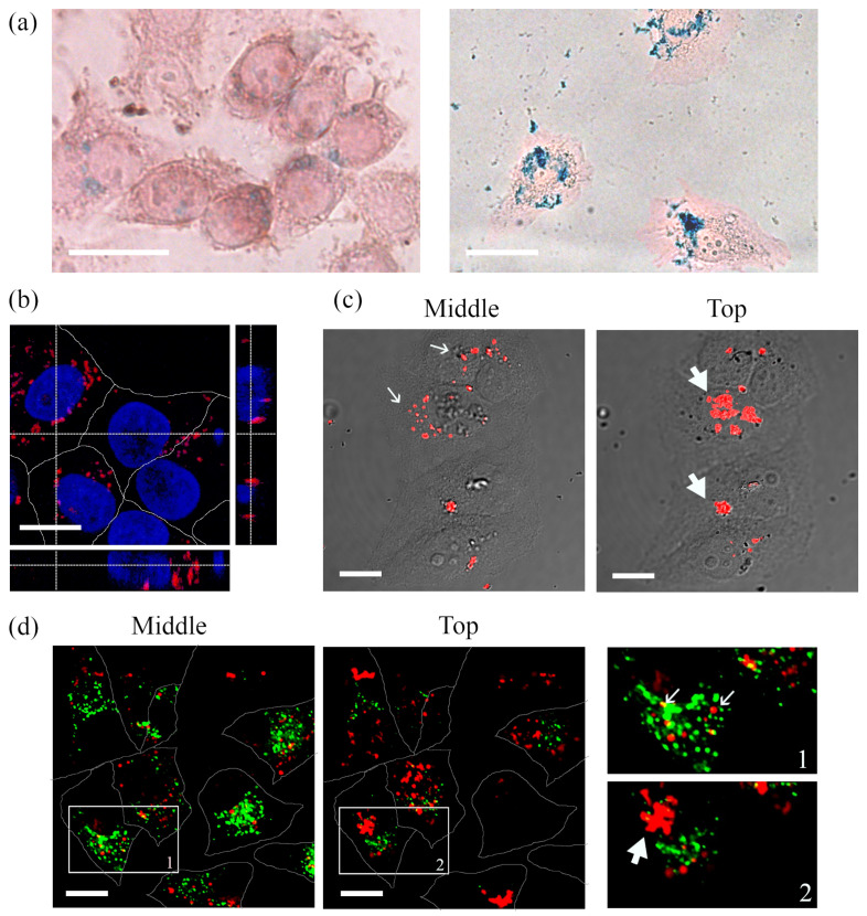
Magnetic-luminescent composites based on semiconductor quantum dots (QDs) and superparamagnetic iron oxide nanoparticles (SPIONs) can serve as a platform combining visualization and therapy. Here, we report the construction of QD-SPION nanocomposites based on synthesized SPIONs and alloyed QDs (CdxZn1-xSeyS1-y)/ZnS solubilized with L-cysteine molecules. The study of the spectral-luminescence characteristics, the kinetics of luminescence decay show the composite's stability in a solution. After incubation with HeLa cells, QDs, SPIONs, and their composites form clusters on the cell surface and associate with endosomes inside the cells. Component-wise analysis of the photoluminescence decay of cell-associated QDs/SPIONs provides information about their localization and aggregate status.
基于半导体量子点 (QD) 和超顺磁性氧化铁纳米粒子 (SPION) 的磁致发光复合材料可用作结合可视化和治疗的平台。在此,我们报告了基于合成的 SPION 和用 L-半胱氨酸分子溶解的合金量子点 (CdxZn1-xSeyS1-y)/ZnS 构建的 QD-SPION 纳米复合材料。对光谱发光特性和发光衰减动力学的研究表明,该复合材料在溶液中具有稳定性。与 HeLa 细胞孵育后,QD、SPION 及其复合材料在细胞表面形成聚集体,并与细胞内的内体结合。与细胞相关的 QD/SPION 光致发光衰减的组分分析提供了有关其定位和聚集状态的信息。