Department of Electrical, Electronic and Systems Engineering, Faculty of Engineering and Built Environment, Universiti Kebangsaan Malaysia, 43600, Bangi, Malaysia.
Department of Computer Science and Engineering, Dhaka University of Engineering & Technology, Gazipur, Gazipur, 1707, Bangladesh.
Sci Rep. 2022 Apr 15;12(1):6319. doi: 10.1038/s41598-022-10309-6.
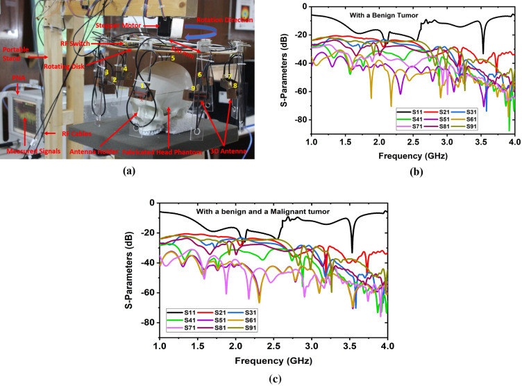
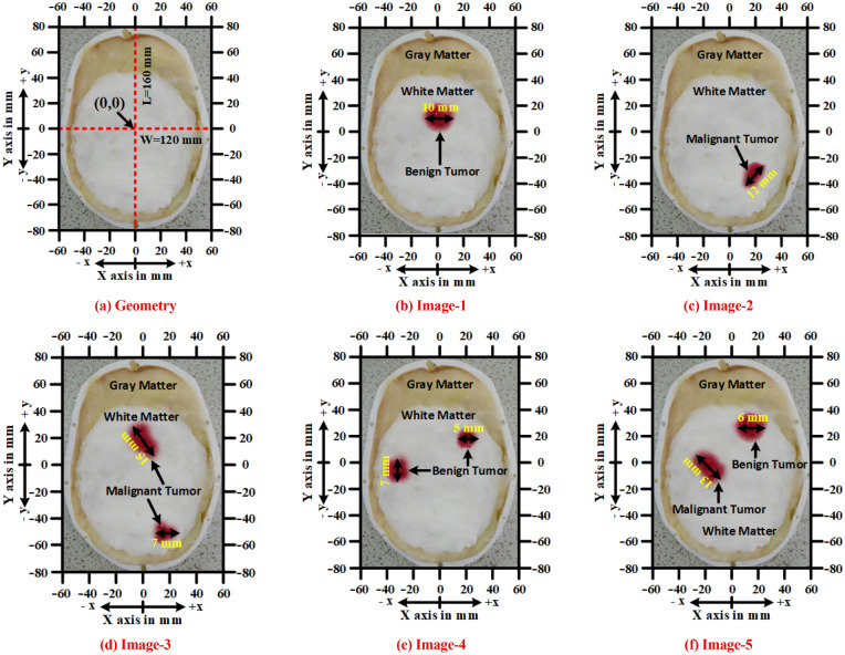
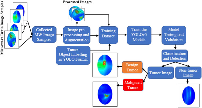
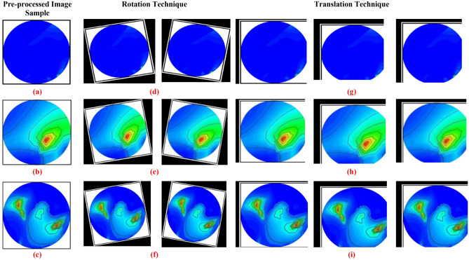
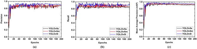
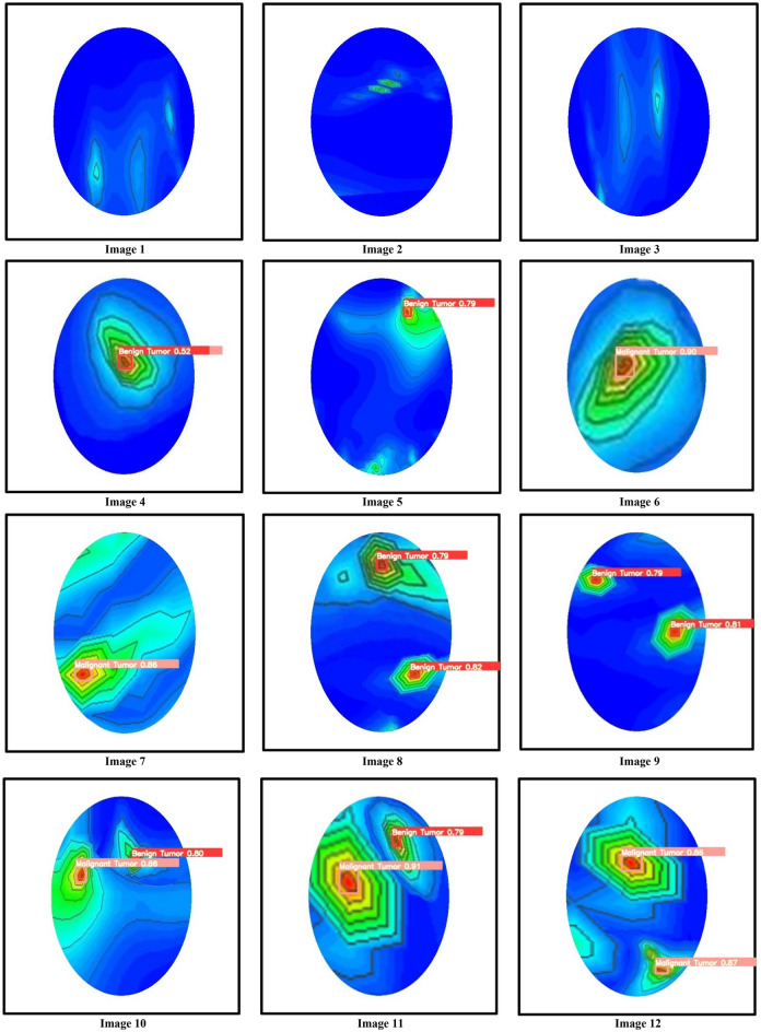
Automated classification and detection of brain abnormalities like a tumor(s) in reconstructed microwave (RMW) brain images are essential for medical application investigation and monitoring disease progression. This paper presents the automatic classification and detection of human brain abnormalities through the deep learning-based YOLOv5 object detection model in a portable microwave head imaging system (MWHI). Initially, four hundred RMW image samples, including non-tumor and tumor(s) in different locations are collected from the implemented MWHI system. The RMW image dimension is 640 × 640 pixels. After that, image pre-processing and augmentation techniques are applied to generate the training dataset, consisting of 4400 images. Later, 80% of images are used to train the models, and 20% are used for testing. Later, from the 80% training dataset, 20% are utilized to validate the models. The detection and classification performances are evaluated by three variations of the YOLOv5 model: YOLOv5s, YOLOv5m, and YOLOv5l. It is investigated that the YOLOv5l model performed better compared to YOLOv5s, YOLOv5m, and state-of-the-art object detection models. The achieved accuracy, precision, sensitivity, specificity, F1-score, mean average precision (mAP), and classification loss are 96.32%, 95.17%, 94.98%, 95.28%, 95.53%, 96.12%, and 0.0130, respectively for the YOLOv5l model. The YOLOv5l model automatically detected tumor(s) accurately with a predicted bounding box including objectness score in RMW images and classified the tumors into benign and malignant classes. So, the YOLOv5l object detection model can be reliable for automatic tumor(s) detection and classification in a portable microwave brain imaging system as a real-time application.
通过基于深度学习的 YOLOv5 对象检测模型,在便携式微波头部成像系统(MWHI)中自动分类和检测人脑异常,是医学应用研究和监测疾病进展的关键。本文提出了在实现的 MWHI 系统中,通过基于深度学习的 YOLOv5 目标检测模型,自动分类和检测人脑异常。最初,从实施的 MWHI 系统中收集了包括不同位置的非肿瘤和肿瘤的 400 个 RMW 图像样本。RMW 图像的维度为 640×640 像素。之后,应用图像预处理和增强技术来生成训练数据集,该数据集由 4400 个图像组成。之后,使用 80%的图像来训练模型,使用 20%的图像来进行测试。之后,从 80%的训练数据集,使用 20%的图像来验证模型。通过 YOLOv5 模型的三个变体:YOLOv5s、YOLOv5m 和 YOLOv5l,评估检测和分类性能。结果表明,YOLOv5l 模型的性能优于 YOLOv5s、YOLOv5m 和最先进的目标检测模型。对于 YOLOv5l 模型,获得的准确性、精度、灵敏度、特异性、F1 分数、平均精度(mAP)和分类损失分别为 96.32%、95.17%、94.98%、95.28%、95.53%、96.12%和 0.0130。YOLOv5l 模型可以在 RMW 图像中自动准确地检测肿瘤,并使用预测的边界框和对象分数对肿瘤进行良性和恶性分类。因此,YOLOv5l 目标检测模型可用于便携式微波脑成像系统中的自动肿瘤检测和分类,作为实时应用。