Department of Ophthalmology, University of Duesseldorf, Moorenstr. 5, D-40225, Düsseldorf, Germany.
Department of Obstetrics and Gynaecology, University of Duesseldorf, Düsseldorf, Germany.
J Med Case Rep. 2022 Apr 21;16(1):167. doi: 10.1186/s13256-022-03369-9.
Retinal arterial occlusive events in young patients are rare. However, because of physiological multifactorial adaptations during pregnancy, retinal vascular occlusive disease may occur spontaneously. In addition, a patent foramen ovale is a risk factor for an ischemic thromboembolic event. Since fluorescein angiography, a central tool in the evaluation of these occlusions, should be avoided during pregnancy, optical coherence tomography angiography, a novel technique, offers a good opportunity for visualizing vascular perfusion of retinal tissue.
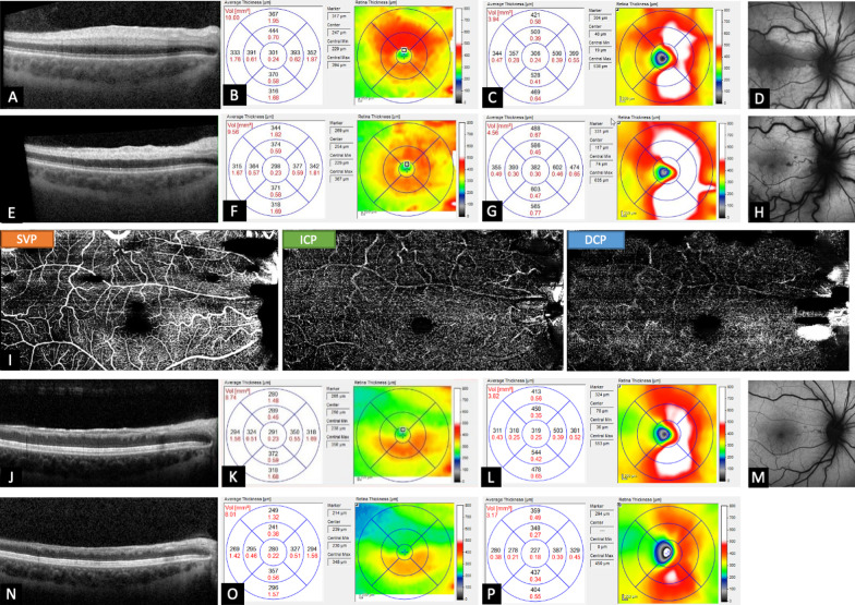
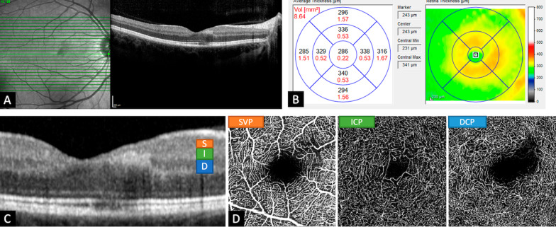
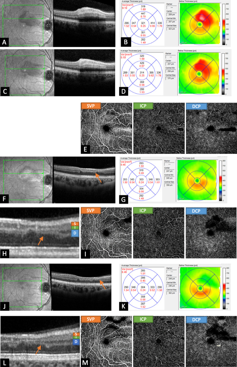
Here we present a case series of three patients (Caucasian, nonsmoker) who visited our clinic owing to acute visual impairment and central scotoma. Using regular optical coherence tomography and optical coherence tomography angiography, retinal vascular occlusions were detected, thus initiating the evaluation of systemic risk factors. We report two patients (30 and 32 years old) who developed cilioretinal artery occlusion but whose etiology differed: one was of thromboembolic origin associated with patent foramen ovale, while the other was caused by hemodynamic blockade secondary to central retinal vein occlusion. In both cases, optical coherence tomography angiography revealed reperfusion of the cilioretinal artery occlusion. However, transient ischemia led to retinal atrophy after a few weeks. In the third patient (32 years old), 8 weeks after onset of scotoma, optical coherence tomography angiography revealed atrophy of the middle layers and impaired perfusion in the deep capillary plexus, and thus a paracentral acute middle maculopathy was diagnosed. All patients regained normal visual acuity and had otherwise uncomplicated pregnancies, and laboratory blood tests did not reveal any defects or alterations.
As shown here, optical coherence tomography angiography enables risk-free imaging of retinal vessel perfusion during pregnancy. Together with regular optical coherence tomography, it allows one to predict functional outcome according to the existing retinal occlusion-related atrophy.
年轻患者发生视网膜动脉阻塞事件较为罕见。然而,由于妊娠期间的生理多因素适应,视网膜血管阻塞性疾病可能会自发发生。此外,卵圆孔未闭是发生缺血性血栓栓塞事件的一个危险因素。由于荧光素血管造影术是评估这些阻塞的核心工具,而在妊娠期间应避免使用,因此新型技术——光学相干断层扫描血管造影术为观察视网膜组织血管灌注提供了良好的机会。
在此,我们报告了三例患者(白种人,不吸烟)的病例系列,他们因急性视力障碍和中央暗点就诊于我们的诊所。通过常规光学相干断层扫描和光学相干断层扫描血管造影术,发现视网膜血管阻塞,从而开始评估系统性危险因素。我们报告了两名(30 岁和 32 岁)发生睫状视网膜动脉阻塞的患者,但病因不同:一位是与卵圆孔未闭相关的血栓栓塞性起源,而另一位是继发于视网膜中央静脉阻塞的血流动力学阻塞。在这两种情况下,光学相干断层扫描血管造影术均显示睫状视网膜动脉阻塞再通。然而,几周后短暂的缺血导致视网膜萎缩。在第三位患者(32 岁)中,在出现暗点 8 周后,光学相干断层扫描血管造影术显示中层萎缩和深层毛细血管丛灌注受损,因此诊断为旁中心急性中黄斑病变。所有患者均恢复正常视力,妊娠均无并发症,实验室血液检查未发现任何缺陷或改变。
如本文所示,光学相干断层扫描血管造影术可在妊娠期间安全地对视网膜血管灌注进行成像。结合常规光学相干断层扫描,它可以根据现有的与视网膜阻塞相关的萎缩来预测功能结果。