Department of Anesthesia, Critical Care, and Pain Medicine, Massachusetts General Hospital, Boston, MA, USA.
The University of Queensland, Brisbane, Australia.
Sci Rep. 2022 Apr 29;12(1):7065. doi: 10.1038/s41598-022-11045-7.
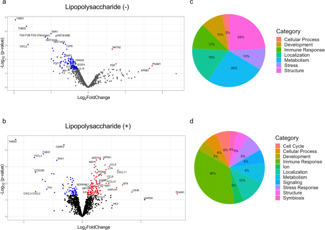
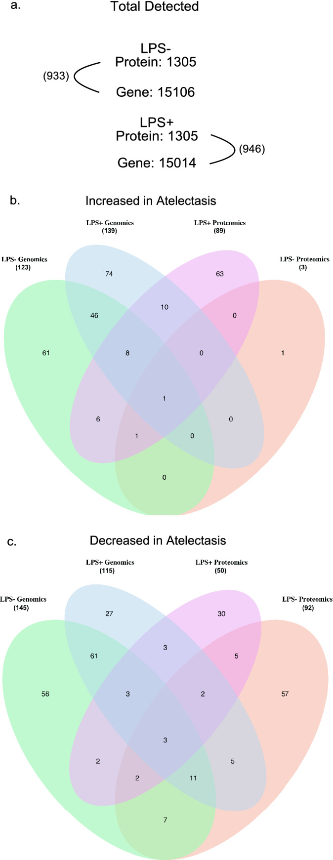
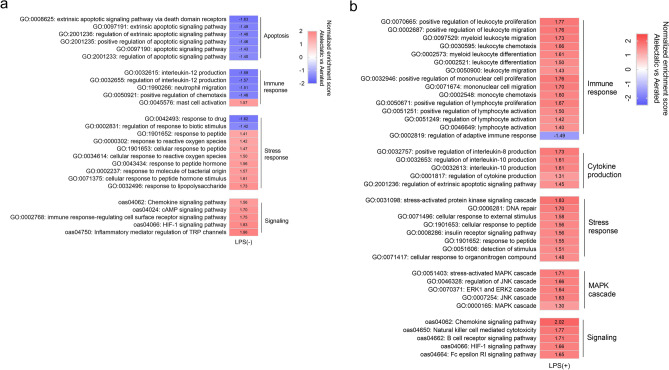
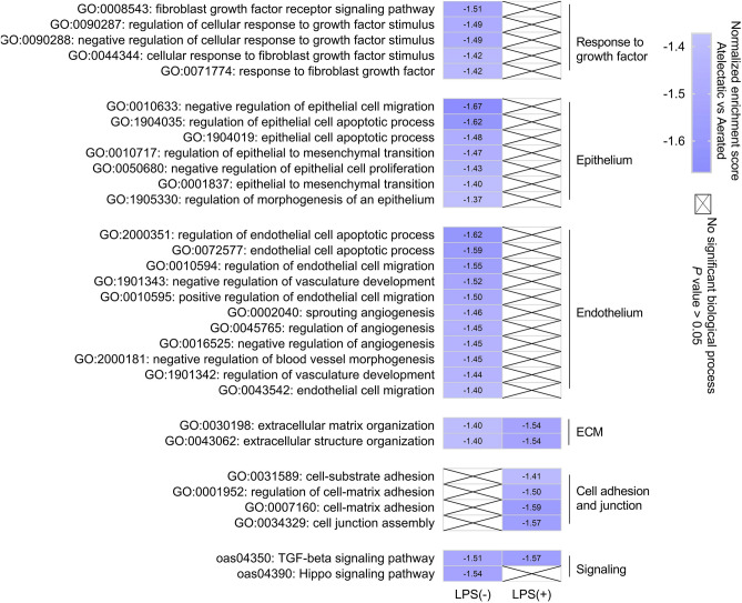
Atelectasis is a frequent clinical condition, yet knowledge is limited and controversial on its biological contribution towards lung injury. We assessed the regional proteomics of atelectatic versus normally-aerated lung tissue to test the hypothesis that immune and alveolar-capillary barrier functions are compromised by purely atelectasis and dysregulated by additional systemic inflammation (lipopolysaccharide, LPS). Without LPS, 130 proteins were differentially abundant in atelectasis versus aerated lung, mostly (n = 126) with less abundance together with negatively enriched processes in immune, endothelial and epithelial function, and Hippo signaling pathway. Instead, LPS-exposed atelectasis produced 174 differentially abundant proteins, mostly (n = 108) increased including acute lung injury marker RAGE and chemokine CCL5. Functional analysis indicated enhanced leukocyte processes and negatively enriched cell-matrix adhesion and cell junction assembly with LPS. Additionally, extracellular matrix organization and TGF-β signaling were negatively enriched in atelectasis with decreased adhesive glycoprotein THBS1 regardless of LPS. Concordance of a subset of transcriptomics and proteomics revealed overlap of leukocyte-related gene-protein pairs and processes. Together, proteomics of exclusively atelectasis indicates decreased immune response, which converts into an increased response with LPS. Alveolar-capillary barrier function-related proteomics response is down-regulated in atelectasis irrespective of LPS. Specific proteomics signatures suggest biological mechanistic and therapeutic targets for atelectasis-associated lung injury.
肺不张是一种常见的临床病症,但人们对其生物学损伤作用的了解有限且存在争议。我们评估了肺不张与正常充气肺组织的局部蛋白质组学,以验证以下假设:单纯肺不张会损害免疫和肺泡毛细血管屏障功能,而全身性炎症(脂多糖,LPS)会使其失调。在没有 LPS 的情况下,肺不张与充气肺相比有 130 种蛋白质丰度存在差异,其中大多数(n=126)丰度降低,同时免疫、内皮和上皮功能以及 Hippo 信号通路的负调控过程也减少。相反,暴露于 LPS 的肺不张会产生 174 种丰度存在差异的蛋白质,其中大多数(n=108)增加,包括急性肺损伤标志物 RAGE 和趋化因子 CCL5。功能分析表明,LPS 会增强白细胞过程,而负调控细胞-基质黏附和细胞连接组装。此外,无论 LPS 如何,细胞外基质组织和 TGF-β信号转导在肺不张中都受到负调控,而细胞黏附糖蛋白 THBS1 减少。转录组学和蛋白质组学的一致性表明,白细胞相关基因-蛋白对和过程存在重叠。总之,单纯性肺不张的蛋白质组学表明免疫反应减弱,而 LPS 会使其反应增强。肺泡毛细血管屏障功能相关的蛋白质组学反应在肺不张中无论 LPS 如何都受到抑制。特定的蛋白质组学特征表明,针对肺不张相关肺损伤的生物学机制和治疗靶点。