• 文献检索
  • 文档翻译
  • 深度研究
  • 学术资讯
  • Suppr Zotero 插件Zotero 插件
  • 邀请有礼
  • 套餐&价格
  • 历史记录
应用&插件
Suppr Zotero 插件Zotero 插件浏览器插件Mac 客户端Windows 客户端微信小程序
定价
高级版会员购买积分包购买API积分包
服务
文献检索文档翻译深度研究API 文档MCP 服务
关于我们
关于 Suppr公司介绍联系我们用户协议隐私条款
关注我们

Suppr 超能文献

核心技术专利:CN118964589B侵权必究
粤ICP备2023148730 号-1Suppr @ 2026

文献检索

告别复杂PubMed语法,用中文像聊天一样搜索,搜遍4000万医学文献。AI智能推荐,让科研检索更轻松。

立即免费搜索

文件翻译

保留排版,准确专业,支持PDF/Word/PPT等文件格式,支持 12+语言互译。

免费翻译文档

深度研究

AI帮你快速写综述,25分钟生成高质量综述,智能提取关键信息,辅助科研写作。

立即免费体验

在乳腺癌鸡胚肿瘤模型中进行体内氟-18 的生产,用于质子治疗范围验证。

In vivo production of fluorine-18 in a chicken egg tumor model of breast cancer for proton therapy range verification.

机构信息

Grupo de Física Nuclear and IPARCOS, Facultad de CC. Físicas, Universidad Complutense de Madrid, CEI Moncloa, 28040, Madrid, Spain.

Instituto de Investigación Sanitaria del Hospital Clínico San Carlos (IdISSC), Ciudad Universitaria, Madrid, Spain.

出版信息

Sci Rep. 2022 Apr 30;12(1):7075. doi: 10.1038/s41598-022-11037-7.

DOI:10.1038/s41598-022-11037-7
PMID:35490180
原文链接:https://pmc.ncbi.nlm.nih.gov/articles/PMC9056503/
Abstract

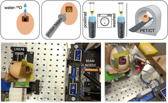
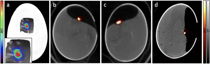
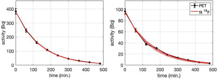
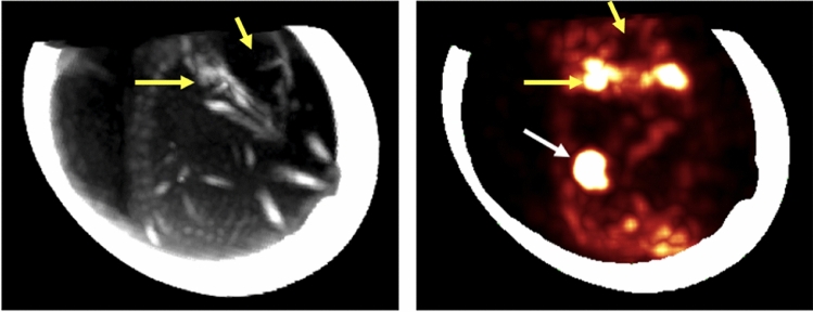
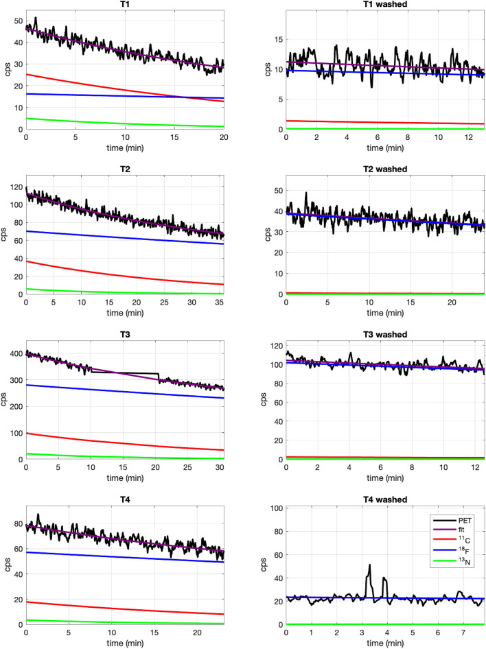
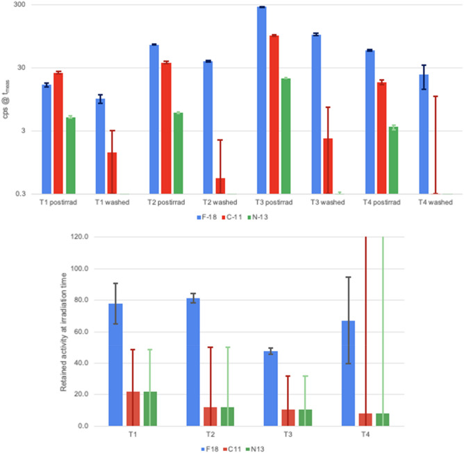
Range verification of clinical protontherapy systems via positron-emission tomography (PET) is not a mature technology, suffering from two major issues: insufficient signal from low-energy protons in the Bragg peak area and biological washout of PET emitters. The use of contrast agents including O, Zn or Cu, isotopes with a high cross section for low-energy protons in nuclear reactions producing PET emitters, has been proposed to enhance the PET signal in the last millimeters of the proton path. However, it remains a challenge to achieve sufficient concentrations of these isotopes in the target volume. Here we investigate the possibilities of O-enriched water (18-W), a potential contrast agent that could be incorporated in large proportions in live tissues by replacing regular water. We hypothesize that 18-W could also mitigate the problem of biological washout, as PET (F) isotopes created inside live cells would remain trapped in the form of fluoride anions (F-), allowing its signal to be detected even hours after irradiation. To test our hypothesis, we designed an experiment with two main goals: first, prove that 18-W can incorporate enough O into a living organism to produce a detectable signal from F after proton irradiation, and second, determine the amount of activity that remains trapped inside the cells. The experiment was performed on a chicken embryo chorioallantoic membrane tumor model of head and neck cancer. Seven eggs with visible tumors were infused with 18-W and irradiated with 8-MeV protons (range in water: 0.74 mm), equivalent to clinical protons at the end of particle range. The activity produced after irradiation was detected and quantified in a small-animal PET-CT scanner, and further studied by placing ex-vivo tumours in a gamma radiation detector. In the acquired images, specific activity of F (originating from 18-W) could be detected in the tumour area of the alive chicken embryo up to 9 h after irradiation, which confirms that low-energy protons can indeed produce a detectable PET signal if a suitable contrast agent is employed. Moreover, dynamic PET studies in two of the eggs evidenced a minimal effect of biological washout, with 68% retained specific F activity at 8 h after irradiation. Furthermore, ex-vivo analysis of 4 irradiated tumours showed that up to 3% of oxygen atoms in the targets were replaced by O from infused 18-W, and evidenced an entrapment of 59% for specific activity of F after washing, supporting our hypothesis that F- ions remain trapped within the cells. An infusion of 18-W can incorporate O in animal tissues by replacing regular water inside cells, producing a PET signal when irradiated with low-energy protons that could be used for range verification in protontherapy. F produced inside cells remains entrapped and suffers from minimal biological washout, allowing for a sharper localization with longer PET acquisitions. Further studies must evaluate the feasibility of this technique in dosimetric conditions closer to clinical practice, in order to define potential protocols for its use in patients.

摘要

临床质子治疗系统的射程验证通过正电子发射断层扫描(PET)不是一项成熟的技术,它存在两个主要问题:在布拉格峰区域内低能质子的信号不足和 PET 发射体的生物洗脱。使用包括 O、Zn 或 Cu 在内的造影剂,这些同位素在核反应中具有与产生 PET 发射体的低能质子的高横截面,被提议用于增强质子路径最后几毫米处的 PET 信号。然而,在靶体积中实现这些同位素的足够浓度仍然是一个挑战。在这里,我们研究了富含氧的水(18-W)的可能性,这是一种潜在的造影剂,可以通过替代普通水来大量掺入活组织中。我们假设 18-W 还可以减轻生物洗脱的问题,因为在活细胞内产生的 PET(F)同位素将以氟化物阴离子(F-)的形式被捕获,即使在辐照后数小时也可以检测到其信号。为了验证我们的假设,我们设计了一个具有两个主要目标的实验:首先,证明 18-W 可以将足够的 O 掺入生物体中,以便在质子辐照后从 F 中产生可检测的信号;其次,确定留在细胞内的活性量。该实验是在头颈部癌症的鸡胚绒毛尿囊膜肿瘤模型上进行的。用 18-W 灌注了七个可见肿瘤的鸡蛋,并以 8-MeV 质子照射(水中射程:0.74 毫米),相当于粒子射程末端的临床质子。用小动物 PET-CT 扫描仪检测和定量辐照后产生的放射性活度,并通过将离体肿瘤置于伽马辐射探测器中进一步研究。在获得的图像中,在辐照后 9 小时,在活着的鸡胚的肿瘤区域可以检测到源自 18-W 的 F 的特异性活度,这证实了如果使用合适的造影剂,低能质子确实可以产生可检测的 PET 信号。此外,两个鸡蛋中的动态 PET 研究表明,生物洗脱的影响很小,辐照后 8 小时保留了 68%的特定 F 活度。此外,对 4 个辐照肿瘤的离体分析表明,靶标中的多达 3%的氧原子被灌注的 18-W 中的 O 取代,并在洗涤后证明 F 的特异性活度有 59%被捕获,这支持了我们的假设,即 F-离子在细胞内被捕获。18-W 的输注可以通过在细胞内用普通水替代来将 O 掺入动物组织中,并用低能质子照射产生 PET 信号,该信号可用于质子治疗中的射程验证。在细胞内产生的 F 仍然被捕获,并且生物洗脱最小化,从而可以通过更长时间的 PET 采集实现更精确的定位。必须进一步研究该技术在更接近临床实践的剂量学条件下的可行性,以便为其在患者中的使用定义潜在方案。

https://cdn.ncbi.nlm.nih.gov/pmc/blobs/1aa5/9056503/11d2dbc751a0/41598_2022_11037_Fig7_HTML.jpg
https://cdn.ncbi.nlm.nih.gov/pmc/blobs/1aa5/9056503/7e113f60ee35/41598_2022_11037_Fig1_HTML.jpg
https://cdn.ncbi.nlm.nih.gov/pmc/blobs/1aa5/9056503/ddecf75b0296/41598_2022_11037_Fig2_HTML.jpg
https://cdn.ncbi.nlm.nih.gov/pmc/blobs/1aa5/9056503/3eb394c31cf4/41598_2022_11037_Fig3_HTML.jpg
https://cdn.ncbi.nlm.nih.gov/pmc/blobs/1aa5/9056503/7e8e8aab6f84/41598_2022_11037_Fig4_HTML.jpg
https://cdn.ncbi.nlm.nih.gov/pmc/blobs/1aa5/9056503/5ef0f0f23210/41598_2022_11037_Fig5_HTML.jpg
https://cdn.ncbi.nlm.nih.gov/pmc/blobs/1aa5/9056503/8cfa0ba99347/41598_2022_11037_Fig6_HTML.jpg
https://cdn.ncbi.nlm.nih.gov/pmc/blobs/1aa5/9056503/11d2dbc751a0/41598_2022_11037_Fig7_HTML.jpg

相似文献

1
In vivo production of fluorine-18 in a chicken egg tumor model of breast cancer for proton therapy range verification.在乳腺癌鸡胚肿瘤模型中进行体内氟-18 的生产,用于质子治疗范围验证。
Sci Rep. 2022 Apr 30;12(1):7075. doi: 10.1038/s41598-022-11037-7.
2
More advantages in detecting bone and soft tissue metastases from prostate cancer using F-PSMA PET/CT.使用F-PSMA PET/CT检测前列腺癌骨和软组织转移方面有更多优势。
Hell J Nucl Med. 2019 Jan-Apr;22(1):6-9. doi: 10.1967/s002449910952. Epub 2019 Mar 7.
3
Feasibility study of using fall-off gradients of early and late PET scans for proton range verification.使用早期和晚期 PET 扫描的下降梯度进行质子射程验证的可行性研究。
Med Phys. 2017 May;44(5):1734-1746. doi: 10.1002/mp.12191. Epub 2017 Mar 30.
4
The production of positron emitters with millisecond half-life during helium beam radiotherapy.在氦束放射治疗中产生具有毫秒半衰期的正电子发射体。
Phys Med Biol. 2019 Dec 5;64(23):235012. doi: 10.1088/1361-6560/ab51c3.
5
Comparing the biological washout of β+-activity induced in mice brain after 12C-ion and proton irradiation.比较12C离子和质子辐照后小鼠大脑中诱导产生的β+活性的生物清除情况。
Phys Med Biol. 2014 Dec 7;59(23):7229-44. doi: 10.1088/0031-9155/59/23/7229. Epub 2014 Nov 10.
6
Feasibility of quasi-prompt PET-based range verification in proton therapy.基于准瞬发正电子发射断层扫描(PET)的质子治疗剂量验证的可行性研究。
Phys Med Biol. 2020 Dec 11;65(24):245013. doi: 10.1088/1361-6560/aba504.
7
Feasibility of proton-activated implantable markers for proton range verification using PET.基于正电子发射断层扫描技术的质子激活植入式标记物在质子射程验证中的可行性研究。
Phys Med Biol. 2013 Nov 7;58(21):7497-512. doi: 10.1088/0031-9155/58/21/7497. Epub 2013 Oct 8.
8
Quantitative assessment of radionuclide production yields in in-beam and offline PET measurements at different proton irradiation facilities.在不同质子辐照设施的在线和离线 PET 测量中对放射性核素生成产额进行定量评估。
Phys Med Biol. 2022 Jul 14;67(15). doi: 10.1088/1361-6560/ac7a89.
9
Monte Carlo patient study on the comparison of prompt gamma and PET imaging for range verification in proton therapy.蒙特卡罗患者研究比较了质子治疗中用于射程验证的瞬发伽马和 PET 成像。
Phys Med Biol. 2011 Feb 21;56(4):1063-82. doi: 10.1088/0031-9155/56/4/012. Epub 2011 Jan 25.
10
First in situ TOF-PET study using digital photon counters for proton range verification.首次使用数字光子计数器进行质子射程验证的原位TOF-PET研究。
Phys Med Biol. 2016 Aug 21;61(16):6203-30. doi: 10.1088/0031-9155/61/16/6203. Epub 2016 Aug 1.

引用本文的文献

1
Zinc-Doped Iron Oxide Nanoparticles as a Proton-Activatable Agent for Dose Range Verification in Proton Therapy.锌掺杂氧化铁纳米颗粒作为质子治疗中剂量范围验证的质子激活剂。
Molecules. 2023 Sep 29;28(19):6874. doi: 10.3390/molecules28196874.
2
Blocking Studies to Evaluate Receptor-Specific Radioligand Binding in the CAM Model by PET and MR Imaging.通过正电子发射断层扫描(PET)和磁共振成像(MR)在鸡胚绒毛尿囊膜(CAM)模型中进行阻断研究以评估受体特异性放射性配体结合
Cancers (Basel). 2022 Aug 10;14(16):3870. doi: 10.3390/cancers14163870.

本文引用的文献

1
Radiochromic film dosimetry for protons up to 10 MeV with EBT2, EBT3 and unlaminated EBT3 films.EBT2、EBT3 及未层压 EBT3 胶片用于能量达 10 MeV 的质子的放射色性胶片剂量测定法。
Phys Med Biol. 2021 May 20;66(11). doi: 10.1088/1361-6560/abfc8d.
2
Feasibility of quasi-prompt PET-based range verification in proton therapy.基于准瞬发正电子发射断层扫描(PET)的质子治疗剂量验证的可行性研究。
Phys Med Biol. 2020 Dec 11;65(24):245013. doi: 10.1088/1361-6560/aba504.
3
Biological washout modelling for in-beam PET: rabbit brain irradiation by C and O ion beams.
用于束流正电子发射断层扫描的生物清除建模:C 和 O 离子束辐照兔脑。
Phys Med Biol. 2020 Jun 1;65(10):105011. doi: 10.1088/1361-6560/ab8532.
4
The Evolving Role of Radiotherapy for Head and Neck Cancer.头颈部癌症放射治疗的演变角色。
Hematol Oncol Clin North Am. 2020 Feb;34(1):91-108. doi: 10.1016/j.hoc.2019.08.019. Epub 2019 Oct 15.
5
Performance Characteristics of the Digital Biograph Vision PET/CT System.Digital Biograph Vision PET/CT 系统的性能特点。
J Nucl Med. 2019 Jul;60(7):1031-1036. doi: 10.2967/jnumed.118.215418. Epub 2019 Jan 10.
6
Performance Evaluation of the Vereos PET/CT System According to the NEMA NU2-2012 Standard.根据 NEMA NU2-2012 标准对 Vereos PET/CT 系统进行性能评估。
J Nucl Med. 2019 Apr;60(4):561-567. doi: 10.2967/jnumed.118.215541. Epub 2018 Oct 25.
7
A full-scale clinical prototype for proton range verification using prompt gamma-ray spectroscopy.基于瞬发伽马射线能谱法的质子射程验证全尺寸临床原型。
Phys Med Biol. 2018 Sep 17;63(18):185019. doi: 10.1088/1361-6560/aad513.
8
Online proton therapy monitoring: clinical test of a Silicon-photodetector-based in-beam PET.在线质子治疗监测:基于硅光电探测器的在线正电子发射断层成像技术的临床测试。
Sci Rep. 2018 Mar 6;8(1):4100. doi: 10.1038/s41598-018-22325-6.
9
Proton Therapy for Head and Neck Cancers.质子治疗头颈部癌症。
Semin Radiat Oncol. 2018 Jan;28(1):53-63. doi: 10.1016/j.semradonc.2017.08.004.
10
Feasibility study of using fall-off gradients of early and late PET scans for proton range verification.使用早期和晚期 PET 扫描的下降梯度进行质子射程验证的可行性研究。
Med Phys. 2017 May;44(5):1734-1746. doi: 10.1002/mp.12191. Epub 2017 Mar 30.