Department of Biological Sciences and Centre for Bioimaging Sciences (CBIS), National University of Singapore (NUS), Singapore, Singapore.
Department of Biological Sciences and Centre for Bioimaging Sciences (CBIS), National University of Singapore (NUS), Singapore, Singapore; School of Chemistry and Molecular Engineering, East China Normal University, Shanghai, China.
J Lipid Res. 2022 Jun;63(6):100220. doi: 10.1016/j.jlr.2022.100220. Epub 2022 Apr 28.
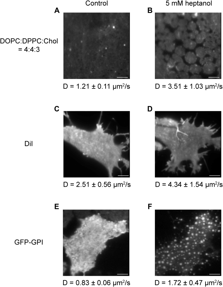
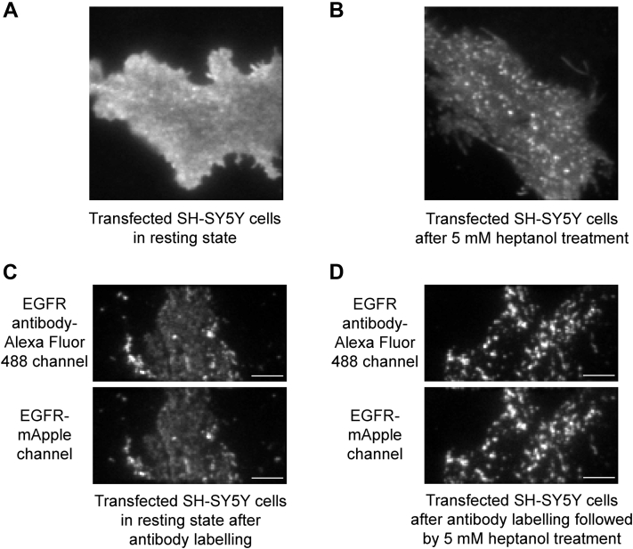
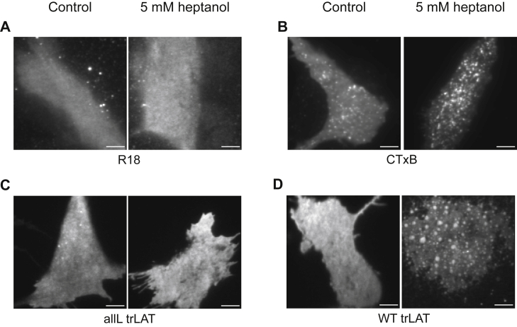
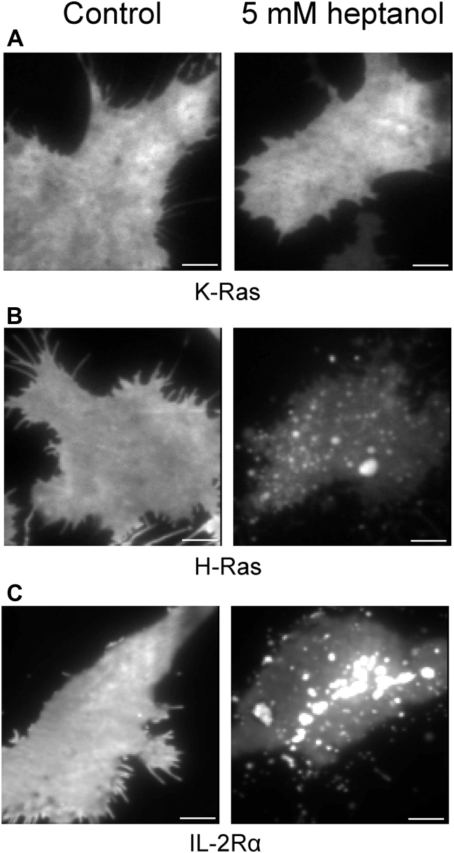
The localization of many membrane proteins within cholesterol- and sphingolipid-containing microdomains is essential for proper cell signaling and function. These membrane domains, however, are too small and dynamic to be recorded, even with modern super-resolution techniques. Therefore, the association of membrane proteins with these domains can only be detected with biochemical assays that destroy the integrity of cells require pooling of many cells and take a long time to perform. Here, we present a simple membrane fluidizer-induced clustering approach to identify the phase-preference of membrane-associated molecules in individual live cells within 10-15 min. Experiments in phase-separated bilayers and live cells on molecules with known phase preference show that heptanol hyperfluidizes the membrane and stabilizes phase separation. This results in a transition from nanosized to micronsized clusters of associated molecules allowing their identification using routine microscopy techniques. Membrane fluidizer-induced clustering is an inexpensive and easy to implement method that can be conducted at large-scale and allows easy identification of protein partitioning in live cell membranes.
许多膜蛋白在含有胆固醇和鞘脂的微域中的定位对于适当的细胞信号转导和功能至关重要。然而,这些膜域太小且动态,即使使用现代超分辨率技术也无法记录。因此,膜蛋白与这些域的关联只能通过生化测定来检测,这些测定会破坏细胞的完整性,需要汇集许多细胞,并且需要很长时间才能完成。在这里,我们提出了一种简单的膜流动性诱导聚集方法,可在 10-15 分钟内鉴定单个活细胞中与膜相关的分子的相偏好。在具有已知相偏好的分子的相分离双层和活细胞中的实验表明,庚醇超流化膜并稳定相分离。这导致相关分子的纳米级簇转变为微米级簇,从而可以使用常规显微镜技术对其进行鉴定。膜流动性诱导聚集是一种廉价且易于实施的方法,可以进行大规模实验,并允许轻松识别活细胞膜中蛋白质的分配。