Xu Li-Na, Yu Xiao-Yu, Chen Wan-Qing, Zhang Song-Mei, Qiu Jing
Department of Oral Implantology, Affiliated Hospital of Stomatology, Nanjing Medical University Nanjing 210029 PR China
Jiangsu Key Laboratory of Oral Disease, Nanjing Medical University Nanjing PR China.
RSC Adv. 2020 Feb 25;10(14):8198-8206. doi: 10.1039/d0ra00154f. eCollection 2020 Feb 24.
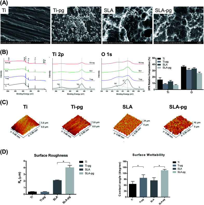
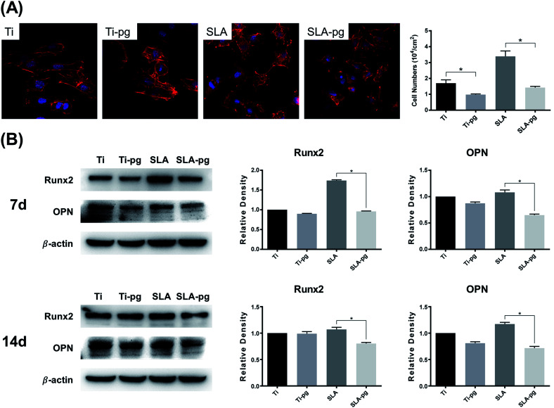
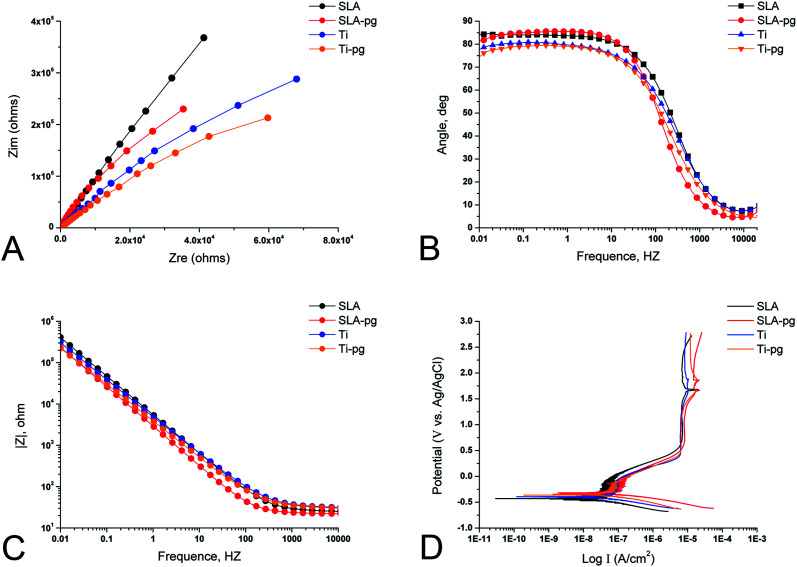
: The study aims to investigate the biocorrosion behavior of on pure and SLA titanium surfaces and its effects on surface characteristics and osteoblast behavior. : Pure and SLA titanium specimens were immersed in culture medium with and incubated for 7 days. colonization on the pure and SLA titanium surfaces was observed by scanning electron microscopy (SEM). The pure and SLA titanium surface characteristics were analyzed X-ray photoelectron spectroscopy (XPS), surface roughness and surface wettability. The corrosion behaviors of pure and SLA titanium specimens were evaluated by electrochemical corrosion test. The osteoblast behavior of MC3T3-E1 cells on the pure and SLA titanium surfaces after colonization was investigated by cell adhesion and western blot assays. : colonized on the pure and SLA titanium surfaces was observed by SEM. The XPS analysis demonstrated reductions in the relative levels of titanium and oxygen and obvious reductions of dominant titanium dioxide (TiO) on both titanium surfaces after immersing the metal in culture. In addition, their roughness and wettability were changed. Correspondingly, the electrochemical corrosion test results revealed significant decreases in the corrosion resistance and increases in the corrosion rate of the pure and SLA titanium specimens after immersion in culture. The results of the study showed that the pre-corroded pure and SLA titanium surfaces by exhibited lower osteocompatibility and down-regulated the adhesion, spreading and osteogenic differentiation abilities of MC3T3-E1 cells. : was able to colonize on the pure and SLA titanium surfaces and weaken their surface properties, especially a decrease in the protective TiO film, which induced the biocorrosion and further negatively affected the osteoblast behavior.
该研究旨在探究[具体物质]在纯钛和酸蚀处理钛(SLA)表面的生物腐蚀行为及其对表面特性和成骨细胞行为的影响。将纯钛和SLA钛标本浸入含有[具体物质]的培养基中并孵育7天。通过扫描电子显微镜(SEM)观察纯钛和SLA钛表面的[具体物质]定植情况。采用X射线光电子能谱(XPS)、表面粗糙度和表面润湿性分析纯钛和SLA钛的表面特性。通过电化学腐蚀试验评估纯钛和SLA钛标本的腐蚀行为。通过细胞黏附试验和蛋白质免疫印迹分析研究MC3T3-E1细胞在[具体物质]定植后在纯钛和SLA钛表面的成骨细胞行为。通过SEM观察到[具体物质]在纯钛和SLA钛表面定植。XPS分析表明,将金属浸入含有[具体物质]的培养基后,两个钛表面的钛和氧相对含量降低,主要的二氧化钛(TiO)明显减少。此外,它们的粗糙度和润湿性发生了变化。相应地,电化学腐蚀试验结果显示,将纯钛和SLA钛标本浸入含有[具体物质]的培养基后,其耐腐蚀性显著降低,腐蚀速率增加。该研究结果表明,经[具体物质]预腐蚀的纯钛和SLA钛表面表现出较低的骨相容性,并下调了MC3T3-E1细胞的黏附、铺展和成骨分化能力。[具体物质]能够在纯钛和SLA钛表面定植并削弱其表面性能,尤其是保护性TiO膜减少,这引发了生物腐蚀并进一步对成骨细胞行为产生负面影响。