• 文献检索
  • 文档翻译
  • 深度研究
  • 学术资讯
  • Suppr Zotero 插件Zotero 插件
  • 邀请有礼
  • 套餐&价格
  • 历史记录
应用&插件
Suppr Zotero 插件Zotero 插件浏览器插件Mac 客户端Windows 客户端微信小程序
定价
高级版会员购买积分包购买API积分包
服务
文献检索文档翻译深度研究API 文档MCP 服务
关于我们
关于 Suppr公司介绍联系我们用户协议隐私条款
关注我们

Suppr 超能文献

核心技术专利:CN118964589B侵权必究
粤ICP备2023148730 号-1Suppr @ 2026

文献检索

告别复杂PubMed语法,用中文像聊天一样搜索,搜遍4000万医学文献。AI智能推荐,让科研检索更轻松。

立即免费搜索

文件翻译

保留排版,准确专业,支持PDF/Word/PPT等文件格式,支持 12+语言互译。

免费翻译文档

深度研究

AI帮你快速写综述,25分钟生成高质量综述,智能提取关键信息,辅助科研写作。

立即免费体验

应用光学相干断层扫描血管造影术分析视网膜前膜眼的血管迂曲。

Vascular tortuosity analysis in eyes with epiretinal membrane imaged by optical coherence tomography angiography.

机构信息

Department of Ophthalmology, Osaka University Graduate School of Medicine, Rm. E7, 2-2 Yamadaoka, Suita, Osaka, 565-0871, Japan.

Integrated Frontier Research for Medical Science Division, Institute for Open and Transdisciplinary Research Initiatives, Osaka University, Suita, Osaka, Japan.

出版信息

BMC Ophthalmol. 2022 May 2;22(1):198. doi: 10.1186/s12886-022-02420-z.

DOI:10.1186/s12886-022-02420-z
PMID:35501767
原文链接:https://pmc.ncbi.nlm.nih.gov/articles/PMC9063110/
Abstract

BACKGROUND

This study aimed to evaluate macular vessel tortuosity using optical coherence tomography angiography (OCTA) and its association with visual outcomes in eyes undergoing surgery for epiretinal membrane (ERM).

METHODS

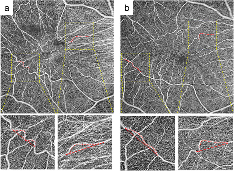
The study included 22 consecutive patients who underwent vitrectomy for ERM between May 2019 and July 2020 and OCTA at Osaka University Hospital. All patients underwent ophthalmologic examinations, including swept-source OCTA. Standard vitrectomy was performed, and the patients were followed up for 6 months postoperatively. Distortion of retinal vessels was calculated using two parameters: the actual vessel length in the vessel section (VL) and the direct vessel branching point distance (BD) in the three quadrants (nasal, temporal, and superior-inferior) of the macula. We analyzed the correlation between these parameters and visual outcomes.

RESULTS

Significantly longer VL was found at 1, 3, and 6 months postoperatively (p = 0.006, 0.008, and 0.022, respectively) in the temporal quadrant compared to baseline temporal VL. Significantly shorter VL was found in nasal quadrants at 1 and 3 months (p = 0.046 and p = 0.018) in the comparison of nasal baseline VL. VL/BDs were correlated with the same postoperative best-corrected visual acuity (BCVA) at 1, 3, and 6 months (p = 0.035, 0.035, and 0.042, respectively) in the superior-inferior quadrant. A significant association of changes in VL and BCVA was found at 3 and 6 months postoperatively in the nasal quadrant (p = 0.018 and 0.0455, respectively).

CONCLUSIONS

Changes in vascular distortion after ERM surgery can be measured using OCTA. The change in vessels around the macula became more linear; this was associated with visual outcomes after surgery.

摘要

背景

本研究旨在通过光学相干断层扫描血管造影术(OCTA)评估黄斑血管迂曲,并评估其与接受外膜切除术(ERM)的眼的视力结果之间的关系。

方法

该研究纳入了 2019 年 5 月至 2020 年 7 月期间在大阪大学医院接受 ERM 玻璃体切除术的 22 例连续患者,并进行了 OCTA 检查。所有患者均接受了眼科检查,包括扫频 OCTA。标准玻璃体切除术,术后随访 6 个月。使用两个参数计算视网膜血管扭曲:血管节段的实际血管长度(VL)和黄斑三个象限(鼻侧、颞侧和上下侧)中直接血管分支点的距离(BD)。我们分析了这些参数与视力结果之间的相关性。

结果

与基线颞侧 VL 相比,术后 1、3 和 6 个月(p=0.006、0.008 和 0.022)颞侧 VL 明显延长。与鼻侧基线 VL 相比,术后 1 和 3 个月(p=0.046 和 p=0.018)鼻侧 VL 明显缩短。VL/BDs 与术后 1、3 和 6 个月的最佳矫正视力(BCVA)呈正相关(p=0.035、0.035 和 0.042),上-下象限。术后 3 个月和 6 个月,鼻侧 VL 和 BCVA 的变化有显著相关性(p=0.018 和 0.0455)。

结论

ERM 手术后血管扭曲的变化可以通过 OCTA 测量。黄斑周围血管的变化变得更加线性;这与手术后的视力结果相关。

https://cdn.ncbi.nlm.nih.gov/pmc/blobs/78c6/9063110/7eafd6050d7e/12886_2022_2420_Fig3_HTML.jpg
https://cdn.ncbi.nlm.nih.gov/pmc/blobs/78c6/9063110/85f1ed397a69/12886_2022_2420_Fig1_HTML.jpg
https://cdn.ncbi.nlm.nih.gov/pmc/blobs/78c6/9063110/cd59b72e0a47/12886_2022_2420_Fig2_HTML.jpg
https://cdn.ncbi.nlm.nih.gov/pmc/blobs/78c6/9063110/7eafd6050d7e/12886_2022_2420_Fig3_HTML.jpg
https://cdn.ncbi.nlm.nih.gov/pmc/blobs/78c6/9063110/85f1ed397a69/12886_2022_2420_Fig1_HTML.jpg
https://cdn.ncbi.nlm.nih.gov/pmc/blobs/78c6/9063110/cd59b72e0a47/12886_2022_2420_Fig2_HTML.jpg
https://cdn.ncbi.nlm.nih.gov/pmc/blobs/78c6/9063110/7eafd6050d7e/12886_2022_2420_Fig3_HTML.jpg

相似文献

1
Vascular tortuosity analysis in eyes with epiretinal membrane imaged by optical coherence tomography angiography.应用光学相干断层扫描血管造影术分析视网膜前膜眼的血管迂曲。
BMC Ophthalmol. 2022 May 2;22(1):198. doi: 10.1186/s12886-022-02420-z.
2
Assessment of macular microvasculature features before and after vitrectomy in the idiopathic macular epiretinal membrane using a grading system: An optical coherence tomography angiography study.采用分级系统评估特发性黄斑视网膜前膜玻璃体切除术前、后黄斑微血管特征:光学相干断层扫描血管造影研究。
Acta Ophthalmol. 2021 Nov;99(7):e1168-e1175. doi: 10.1111/aos.14753. Epub 2021 Jan 10.
3
Macular capillary plexuses after epiretinal membrane surgery: an optical coherence tomography angiography study.视网膜膜后手术的黄斑毛细血管丛:光学相干断层扫描血管造影研究。
Br J Ophthalmol. 2018 Aug;102(8):1086-1091. doi: 10.1136/bjophthalmol-2017-311188. Epub 2017 Oct 31.
4
Quantitative analysis of macular contraction in idiopathic epiretinal membrane.特发性视网膜前膜黄斑收缩的定量分析。
BMC Ophthalmol. 2014 Apr 16;14:51. doi: 10.1186/1471-2415-14-51.
5
Comparative analysis of metamorphopsia and aniseikonia after vitrectomy for epiretinal membrane, macular hole, or rhegmatogenous retinal detachment.比较分析增生性玻璃体视网膜病变、黄斑裂孔或孔源性视网膜脱离玻璃体切除术后的形觉扭曲和像差。
PLoS One. 2020 May 8;15(5):e0232758. doi: 10.1371/journal.pone.0232758. eCollection 2020.
6
[Correlation of capillary plexus with visual acuity in idiopathic macular epiretinal membrane eyes using optical coherence tomography angiography].[利用光学相干断层扫描血管造影术研究特发性黄斑视网膜前膜眼中毛细血管丛与视力的相关性]
Zhonghua Yan Ke Za Zhi. 2019 Oct 11;55(10):757-762. doi: 10.3760/cma.j.issn.0412-4081.2019.10.006.
7
Associations between macular findings by optical coherence tomography and visual outcomes after epiretinal membrane removal.光学相干断层扫描黄斑区表现与视网膜前膜去除术后视觉预后的相关性。
Am J Ophthalmol. 2009 Mar;147(3):473-480.e3. doi: 10.1016/j.ajo.2008.09.020. Epub 2008 Dec 3.
8
Peripapillary RNFL Thickness Changes Evaluated with Spectral Domain Optical Coherence Tomography after Uncomplicated Macular Surgery for Epiretinal Membrane.视网膜前膜单纯性黄斑手术后通过光谱域光学相干断层扫描评估的视乳头周围视网膜神经纤维层厚度变化
Semin Ophthalmol. 2017;32(4):449-455. doi: 10.3109/08820538.2015.1119858. Epub 2016 Apr 14.
9
A study analyzing macular microvasculature features after vitrectomy using OCT angiography in patients with idiopathic macular epiretinal membrane.一项使用 OCT 血管造影术分析特发性黄斑视网膜前膜患者玻璃体切除术后黄斑微血管特征的研究。
BMC Ophthalmol. 2020 Apr 22;20(1):165. doi: 10.1186/s12886-020-01429-6.
10
Relationship between the morphology of the foveal avascular zone and the degree of aniseikonia before and after vitrectomy in patients with unilateral epiretinal membrane.单侧视网膜前膜患者玻璃体切除术前、后黄斑无血管区形态与两眼物象不等程度的关系
Graefes Arch Clin Exp Ophthalmol. 2019 Mar;257(3):507-515. doi: 10.1007/s00417-019-04245-x. Epub 2019 Jan 14.

引用本文的文献

1
Changes in retinal vascular geometry after vitrectomy in idiopathic and uveitic epiretinal membrane.特发性和葡萄膜炎性视网膜前膜玻璃体切除术后视网膜血管几何形态的变化
Sci Rep. 2025 Jul 1;15(1):21415. doi: 10.1038/s41598-025-06771-7.
2
Radiomic Feature Extraction from OCT Angiography of Idiopathic Epiretinal Membranes and Correlation with Visual Acuity: A Pilot Study.特发性视网膜前膜光学相干断层扫描血管造影的影像组学特征提取及其与视力的相关性:一项初步研究
Ophthalmol Sci. 2025 Jan 21;5(3):100716. doi: 10.1016/j.xops.2025.100716. eCollection 2025 May-Jun.
3
Topographic correlation of microperimetry with foveal microstructure characteristics in idiopathic epiretinal membrane patients with an ectopic inner foveal layer.

本文引用的文献

1
Association between displacement and thickness of macula after vitrectomy in eyes with epiretinal membrane.玻璃体切除术后眼内视网膜前膜的黄斑位移与厚度的关系。
Sci Rep. 2020 Aug 6;10(1):13227. doi: 10.1038/s41598-020-70197-6.
2
Disorganization of Retinal Inner Layers as a Biomarker for Idiopathic Epiretinal Membrane After Macular Surgery-The DREAM Study.视网膜内层层状紊乱作为黄斑手术后特发性视网膜前膜的生物标志物:DREAM 研究。
Am J Ophthalmol. 2018 Dec;196:129-135. doi: 10.1016/j.ajo.2018.08.037. Epub 2018 Sep 1.
3
Inner Nuclear Layer Thickness, a Biomarker of Metamorphopsia in Epiretinal Membrane, Correlates With Tangential Retinal Displacement.
特发性黄斑前膜伴黄斑中心凹内层异位患者微视野检查与黄斑中心凹微观结构特征的地形图相关性研究
Eye Vis (Lond). 2025 Feb 1;12(1):4. doi: 10.1186/s40662-024-00419-9.
4
Assessment of clinical outcomes and prognostic factors following membrane peeling in idiopathic epiretinal membrane using EIFL staging system: an optical coherence tomography angiography analysis.使用EIFL分期系统评估特发性视网膜前膜剥膜术后的临床结局和预后因素:光学相干断层扫描血管造影分析
BMC Ophthalmol. 2025 Jan 30;25(1):54. doi: 10.1186/s12886-025-03889-0.
5
Location and Extent of Paravascular Nerve Fiber Layer Clefts in Eyes with Epiretinal Membranes.视网膜前膜患者眼内血管旁神经纤维层裂孔的位置及范围
J Clin Med. 2024 Sep 26;13(19):5731. doi: 10.3390/jcm13195731.
6
Microvascular Changes after Epiretinal Membrane Vitrectomy with Intraoperative Intravitreal Dexamethasone Implant: An OCT Angiography Analysis.术中玻璃体腔内植入地塞米松的视网膜前膜玻璃体切除术后的微血管变化:一项光学相干断层扫描血管造影分析
Diagnostics (Basel). 2024 Feb 13;14(4):411. doi: 10.3390/diagnostics14040411.
7
Quantitative assessment of retinal microvascular remodeling in eyes that underwent idiopathic epiretinal membrane surgery.对接受特发性视网膜前膜手术的眼睛的视网膜微血管重塑进行定量评估。
Front Cell Dev Biol. 2023 Apr 20;11:1164529. doi: 10.3389/fcell.2023.1164529. eCollection 2023.
8
Retinal Vascular Tortuosity Index Change after Idiopathic Epiretinal Membrane Surgery: Does Internal Limiting Membrane Peeling Affect Retinal Vascular Tortuosity?特发性视网膜前膜手术后视网膜血管迂曲指数的变化:内界膜剥除术是否会影响视网膜血管迂曲?
Diagnostics (Basel). 2023 Feb 20;13(4):797. doi: 10.3390/diagnostics13040797.
内核层厚度,一种与视网膜前膜导致的变形有关的生物标志物,与切线性视网膜移位相关。
Am J Ophthalmol. 2018 Sep;193:20-27. doi: 10.1016/j.ajo.2018.06.001. Epub 2018 Jun 8.
4
Insights Into Epiretinal Membranes: Presence of Ectopic Inner Foveal Layers and a New Optical Coherence Tomography Staging Scheme.视网膜前膜的见解:异位中央凹内层的存在及一种新的光学相干断层扫描分期方案
Am J Ophthalmol. 2017 Mar;175:99-113. doi: 10.1016/j.ajo.2016.12.006. Epub 2016 Dec 18.
5
MEASUREMENT OF RETINAL DISPLACEMENT AND METAMORPHOPSIA AFTER EPIRETINAL MEMBRANE OR MACULAR HOLE SURGERY.视网膜前膜或黄斑裂孔手术后视网膜移位和视物变形的测量
Retina. 2016 Apr;36(4):695-702. doi: 10.1097/IAE.0000000000000768.
6
Quantitative analysis of macular contraction in idiopathic epiretinal membrane.特发性视网膜前膜黄斑收缩的定量分析。
BMC Ophthalmol. 2014 Apr 16;14:51. doi: 10.1186/1471-2415-14-51.
7
Quantification of changes in metamorphopsia and retinal contraction in eyes with spontaneous separation of idiopathic epiretinal membrane.定量分析特发性眼内细胞层视网膜前膜自发分离后出现的形觉剥夺性变化和视网膜收缩。
Eye (Lond). 2013 Aug;27(8):924-30. doi: 10.1038/eye.2013.108. Epub 2013 May 31.
8
Quantification of retinal tangential movement in epiretinal membranes.视网膜前膜中视网膜切向运动的定量分析。
Ophthalmology. 2012 Sep;119(9):1886-91. doi: 10.1016/j.ophtha.2012.03.022. Epub 2012 May 26.
9
Time course of changes in metamorphopsia, visual acuity, and OCT parameters after successful epiretinal membrane surgery.成功的视网膜内膜手术后,像差、视力和 OCT 参数变化的时间过程。
Invest Ophthalmol Vis Sci. 2012 Jun 14;53(7):3592-7. doi: 10.1167/iovs.12-9493.
10
Improvement of horizontal macular contraction after surgical removal of epiretinal membranes.手术切除视网膜内界膜后水平性黄斑皱缩的改善。
Eye (Lond). 2011 Jun;25(6):754-61. doi: 10.1038/eye.2011.48. Epub 2011 Apr 1.