Center for Infectious Diseases and Pathogen Biology, Institute of Virology and AIDS Research, Key Laboratory of Organ Regeneration and Transplantation of the Ministry of Education, The First Hospital of Jilin University, Jilin, 130021, China.
Center for Infectious Diseases and Pathogen Biology, Institute of Virology and AIDS Research, Key Laboratory of Organ Regeneration and Transplantation of the Ministry of Education, The First Hospital of Jilin University, Jilin, 130021, China.
Virol Sin. 2022 Jun;37(3):418-426. doi: 10.1016/j.virs.2022.04.013. Epub 2022 May 3.
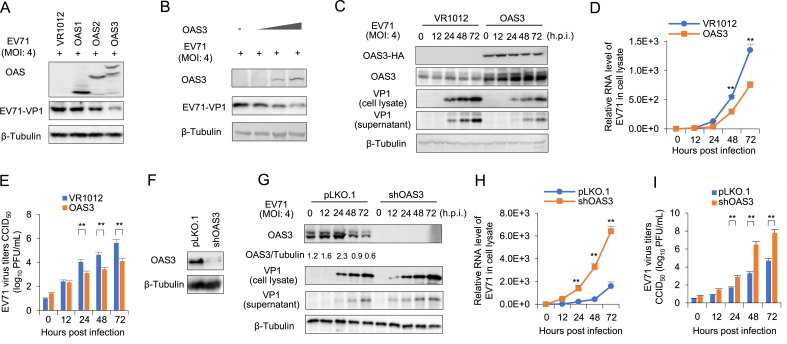
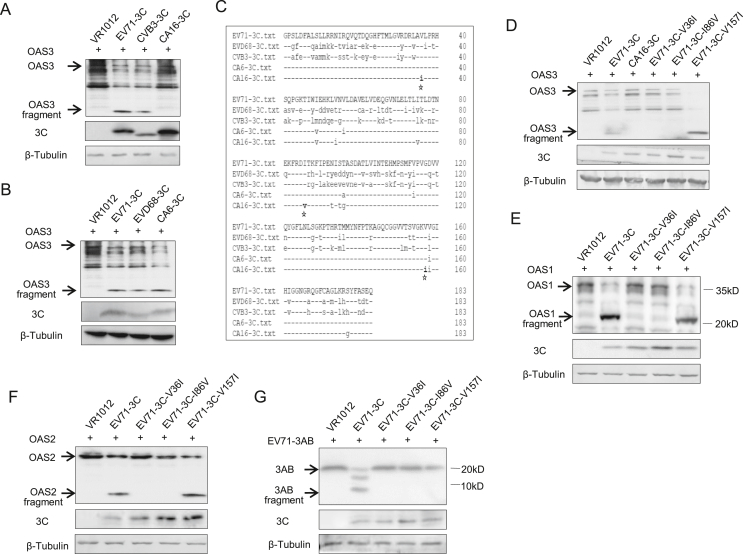
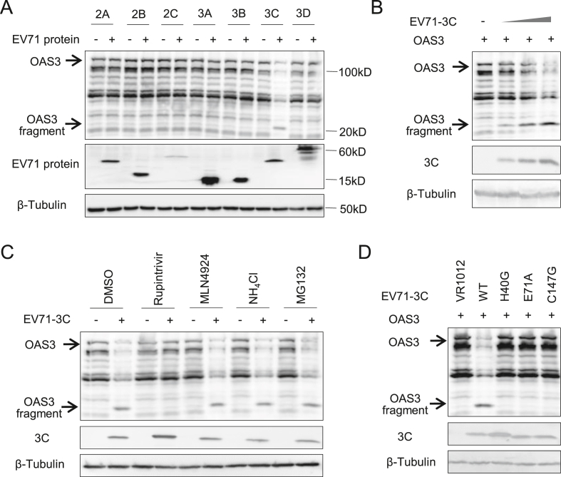
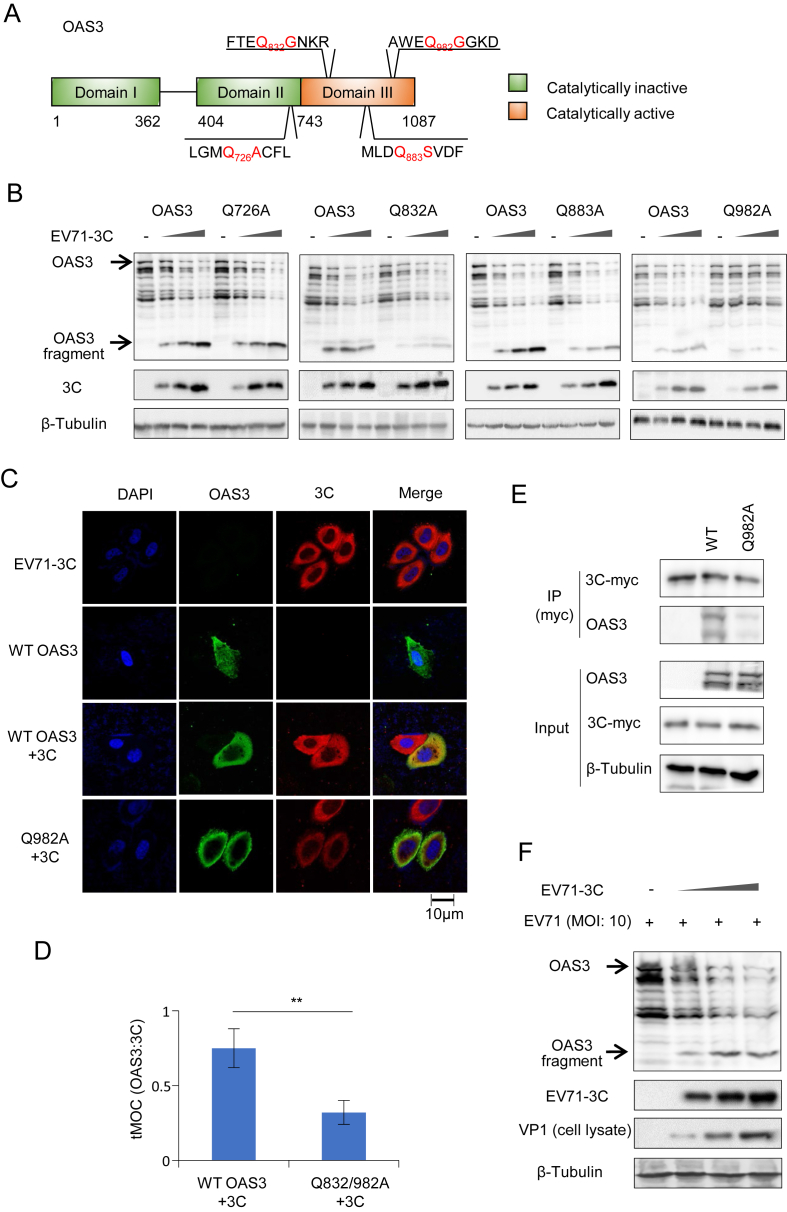
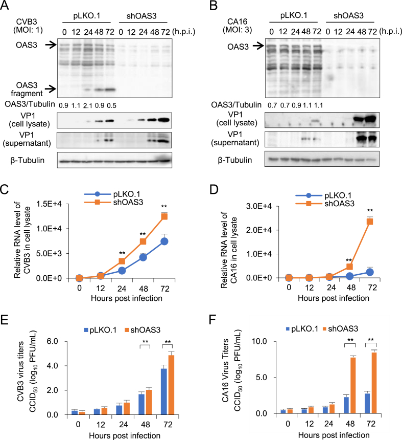
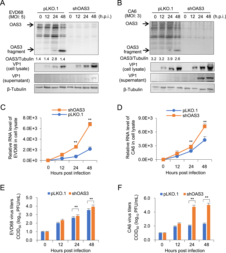
The global spread of enteroviruses (EVs) has become more frequent, severe and life-threatening. Intereron (IFN) I has been proved to control EVs by regulating IFN-stimulated genes (ISG) expression. 2'-5'-oligoadenylate synthetases 3 (OAS3) is an important ISG in the OAS/RNase L antiviral system. The relationship between OAS3 and EVs is still unclear. Here, we reveal that OAS3, superior to OAS1 and OAS2, significantly inhibited EV71 replication in vitro. However, EV71 utilized autologous 3C protease (3C) to cleave intracellular OAS3 and enhance viral replication. Rupintrivir, a human rhinovirus 3C protease inhibitor, completely abolished the cleavage of EV71 3C on OAS3. And the proteolytically deficient mutants H40G, E71A, and C147G of EV71 3C also lost the ability of OAS3 cleavage. Mechanistically, the Q982-G983 motif in C-terminal of OAS3 was identified as a crucial 3C cutting site. Further investigation indicated that OAS3 inhibited not only EV71 but also Coxsackievirus B3 (CVB3), Coxsackievirus A16 (CA16), Enterovirus D68 (EVD68), and Coxsackievirus A6 (CA6) subtypes. Notably, unlike other four subtypes, CA16 3C could not cleave OAS3. Two key amino acids variation Ile36 and Val86 in CA16 3C might result in weak and delayed virus replication of CA16 because of failure of OAS and 3AB cleavage. Our works elucidate the broad anti-EVs function of OAS3, and illuminate a novel mechanism by which EV71 use 3C to escape the antiviral effect of OAS3. These findings can be an important entry point for developing novel therapeutic strategies for multiple EVs infection.
肠道病毒(EVs)在全球范围内的传播变得更加频繁、严重和危及生命。干扰素(IFN)I 已被证明通过调节干扰素刺激基因(ISG)表达来控制 EVs。2′-5′寡聚腺苷酸合成酶 3(OAS3)是 OAS/RNase L 抗病毒系统中的重要 ISG。OAS3 与 EVs 之间的关系尚不清楚。在这里,我们揭示 OAS3,优于 OAS1 和 OAS2,可显著抑制 EV71 在体外的复制。然而,EV71 利用自身的 3C 蛋白酶(3C)切割细胞内的 OAS3,从而增强病毒复制。Rupintrivir,一种人鼻病毒 3C 蛋白酶抑制剂,完全阻止了 EV71 3C 对 OAS3 的切割。并且 EV71 3C 的 H40G、E71A 和 C147G 等无酶切活性的突变体也失去了对 OAS3 切割的能力。从机制上讲,OAS3 C 端的 Q982-G983 基序被鉴定为一个关键的 3C 切割位点。进一步的研究表明,OAS3 不仅抑制 EV71,还抑制柯萨奇病毒 B3(CVB3)、柯萨奇病毒 A16(CA16)、肠道病毒 D68(EVD68)和柯萨奇病毒 A6(CA6)亚型。值得注意的是,与其他四种亚型不同,CA16 3C 不能切割 OAS3。CA16 3C 中的两个关键氨基酸变异 Ile36 和 Val86 可能导致 CA16 病毒复制减弱和延迟,因为 OAS 和 3AB 的切割失败。我们的工作阐明了 OAS3 对多种 EVs 的广泛抗病毒功能,并阐明了 EV71 利用 3C 逃避 OAS3 抗病毒作用的新机制。这些发现可以为开发针对多种 EV 感染的新型治疗策略提供重要切入点。