• 文献检索
  • 文档翻译
  • 深度研究
  • 学术资讯
  • Suppr Zotero 插件Zotero 插件
  • 邀请有礼
  • 套餐&价格
  • 历史记录
应用&插件
Suppr Zotero 插件Zotero 插件浏览器插件Mac 客户端Windows 客户端微信小程序
定价
高级版会员购买积分包购买API积分包
服务
文献检索文档翻译深度研究API 文档MCP 服务
关于我们
关于 Suppr公司介绍联系我们用户协议隐私条款
关注我们

Suppr 超能文献

核心技术专利:CN118964589B侵权必究
粤ICP备2023148730 号-1Suppr @ 2026

文献检索

告别复杂PubMed语法,用中文像聊天一样搜索,搜遍4000万医学文献。AI智能推荐,让科研检索更轻松。

立即免费搜索

文件翻译

保留排版,准确专业,支持PDF/Word/PPT等文件格式,支持 12+语言互译。

免费翻译文档

深度研究

AI帮你快速写综述,25分钟生成高质量综述,智能提取关键信息,辅助科研写作。

立即免费体验

具有九倍膨胀(NIFS)水凝胶的扩展显微镜技术可在传统显微镜上进行细胞超微结构成像。

Expansion microscopy with ninefold swelling (NIFS) hydrogel permits cellular ultrastructure imaging on conventional microscope.

作者信息

Li Hongxia, Warden Antony R, He Jie, Shen Guangxia, Ding Xianting

机构信息

State Key Laboratory of Oncogenes and Related Genes, Institute for Personalized Medicine, School of Biomedical Engineering, Shanghai Jiao Tong University, Shanghai 200030, China.

出版信息

Sci Adv. 2022 May 6;8(18):eabm4006. doi: 10.1126/sciadv.abm4006. Epub 2022 May 4.

DOI:10.1126/sciadv.abm4006
PMID:35507653
原文链接:https://pmc.ncbi.nlm.nih.gov/articles/PMC9067917/
Abstract

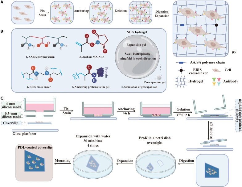
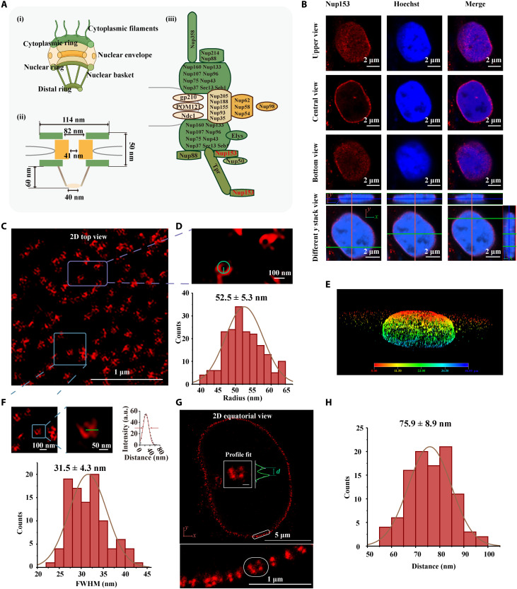
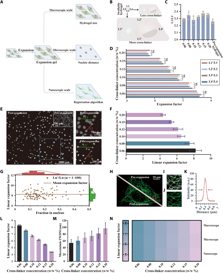
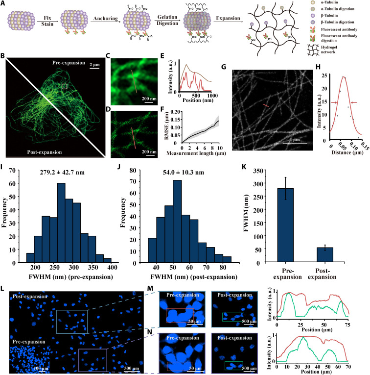
Superresolution microscopy enables probing of cellular ultrastructures. However, its widespread applications are limited by the need for expensive machinery, specific hardware, and sophisticated data processing. Expansion microscopy (ExM) improves the resolution of conventional microscopy by physically expanding biological specimens before imaging and currently provides 70-nm resolution, which still lags behind that of modern superresolution microscopy (30 nm). Here, we demonstrate a ninefold swelling (NIFS) hydrogel, that can reduce ExM resolution to 31 nm when using regular traditional microscopy. We also design a detachable chip that integrates all the experimental operations to facilitate the maximal reproducibility of this high-resolution imaging technology. We demonstrate this technique on the superimaging of nuclear pore complex and clathrin-coated pits, whose structures can hardly be resolved by conventional microscopy. The method presented here offers a universal platform with superresolution imaging to unveil cellular ultrastructural details using standard conventional laboratory microscopes.

摘要

超分辨率显微镜能够探测细胞超微结构。然而,其广泛应用受到昂贵设备、特定硬件和复杂数据处理需求的限制。扩展显微镜(ExM)通过在成像前对生物样本进行物理扩展来提高传统显微镜的分辨率,目前可提供约70纳米的分辨率,仍落后于现代超分辨率显微镜(约30纳米)。在此,我们展示了一种九倍膨胀(NIFS)水凝胶,使用常规传统显微镜时,它可将ExM分辨率降低至31纳米。我们还设计了一种可拆卸芯片,集成了所有实验操作,以促进这种高分辨率成像技术的最大可重复性。我们在核孔复合体和网格蛋白包被小窝的超成像上展示了这项技术,其结构很难通过传统显微镜分辨。本文提出的方法提供了一个具有超分辨率成像的通用平台,可使用标准的常规实验室显微镜揭示细胞超微结构细节。

https://cdn.ncbi.nlm.nih.gov/pmc/blobs/2fc8/9067917/5e1f4956551f/sciadv.abm4006-f4.jpg
https://cdn.ncbi.nlm.nih.gov/pmc/blobs/2fc8/9067917/5d402bb998e9/sciadv.abm4006-f1.jpg
https://cdn.ncbi.nlm.nih.gov/pmc/blobs/2fc8/9067917/eb17d9e88ce9/sciadv.abm4006-f2.jpg
https://cdn.ncbi.nlm.nih.gov/pmc/blobs/2fc8/9067917/1fae58e01709/sciadv.abm4006-f3.jpg
https://cdn.ncbi.nlm.nih.gov/pmc/blobs/2fc8/9067917/5e1f4956551f/sciadv.abm4006-f4.jpg

相似文献

1
Expansion microscopy with ninefold swelling (NIFS) hydrogel permits cellular ultrastructure imaging on conventional microscope.具有九倍膨胀(NIFS)水凝胶的扩展显微镜技术可在传统显微镜上进行细胞超微结构成像。
Sci Adv. 2022 May 6;8(18):eabm4006. doi: 10.1126/sciadv.abm4006. Epub 2022 May 4.
2
Practical method for superresolution imaging of primary cilia and centrioles by expansion microscopy using an amplibody for fluorescence signal amplification.利用用于荧光信号放大的抗体进行扩展显微镜超分辨成像,实现初级纤毛和中心体的实用方法。
Mol Biol Cell. 2020 Sep 15;31(20):2195-2206. doi: 10.1091/mbc.E20-04-0250. Epub 2020 Jul 29.
3
X10 expansion microscopy enables 25-nm resolution on conventional microscopes.X10 扩展显微镜可在常规显微镜上实现 25nm 的分辨率。
EMBO Rep. 2018 Sep;19(9). doi: 10.15252/embr.201845836. Epub 2018 Jul 9.
4
Superresolution imaging of Drosophila tissues using expansion microscopy.使用扩展显微镜对果蝇组织进行超分辨率成像。
Mol Biol Cell. 2018 Jun 15;29(12):1413-1421. doi: 10.1091/mbc.E17-10-0583. Epub 2018 Apr 24.
5
Expanding boundaries - a cell biologist's guide to expansion microscopy.拓展边界——细胞生物学家的扩展显微镜指南。
J Cell Sci. 2024 Apr 1;137(7). doi: 10.1242/jcs.260765. Epub 2024 Apr 17.
6
Expansion Mini-Microscopy: An Enabling Alternative in Point-of-Care Diagnostics.扩展式微型显微镜:即时诊断中的一种可行替代方法。
Curr Opin Biomed Eng. 2017 Mar;1:45-53. doi: 10.1016/j.cobme.2017.03.001. Epub 2017 Mar 22.
7
Expansion Microscopy with Multifunctional Polymer Dots.多功能聚合物点的扩展显微镜技术。
Adv Mater. 2021 Jun;33(25):e2007854. doi: 10.1002/adma.202007854. Epub 2021 May 14.
8
Optical imaging. Expansion microscopy.光学成像。扩展显微镜技术。
Science. 2015 Jan 30;347(6221):543-8. doi: 10.1126/science.1260088. Epub 2015 Jan 15.
9
Ultrastructure expansion microscopy (U-ExM).超微结构扩展显微镜(U-ExM)。
Methods Cell Biol. 2021;161:57-81. doi: 10.1016/bs.mcb.2020.05.006. Epub 2020 Jul 1.
10
Expansion Microscopy for Beginners: Visualizing Microtubules in Expanded Cultured HeLa Cells.初学者的扩展显微镜技术:在扩展培养的 HeLa 细胞中可视化微管。
Curr Protoc Neurosci. 2020 Jun;92(1):e96. doi: 10.1002/cpns.96.

引用本文的文献

1
Single extracellular vesicle imaging via rolling circle amplification-expansion microscopy.通过滚环扩增-扩张显微镜对单个细胞外囊泡进行成像。
Nat Commun. 2025 Aug 13;16(1):7498. doi: 10.1038/s41467-025-62613-0.
2
Subdiffraction Imaging of Cleared and Expanded Large-Scale Tissues.清除和扩展后的大规模组织的亚衍射成像
Chem Biomed Imaging. 2024 Jun 18;2(8):542-559. doi: 10.1021/cbmi.4c00013. eCollection 2024 Aug 26.
3
Expanding boundaries - a cell biologist's guide to expansion microscopy.拓展边界——细胞生物学家的扩展显微镜指南。

本文引用的文献

1
Toward High Spatially Resolved Proteomics Using Expansion Microscopy.利用扩展显微镜实现高空间分辨率蛋白质组学
Anal Chem. 2021 Sep 14;93(36):12195-12203. doi: 10.1021/acs.analchem.0c05372. Epub 2021 Aug 27.
2
A Universal Labeling Strategy for Nucleic Acids in Expansion Microscopy.用于扩展显微镜的核酸通用标记策略。
J Am Chem Soc. 2021 Sep 1;143(34):13782-13789. doi: 10.1021/jacs.1c05931. Epub 2021 Aug 23.
3
Expansion Microscopy with Multifunctional Polymer Dots.多功能聚合物点的扩展显微镜技术。
J Cell Sci. 2024 Apr 1;137(7). doi: 10.1242/jcs.260765. Epub 2024 Apr 17.
4
Label-Retention Expansion Microscopy (LR-ExM) for Enhanced Fluorescent Signals using Trifunctional Probes.标签保留扩展显微镜(LR-ExM)技术用于三功能探针增强荧光信号。
Curr Protoc. 2024 Jan;4(1):e973. doi: 10.1002/cpz1.973.
5
GelMap: intrinsic calibration and deformation mapping for expansion microscopy.凝胶图:膨胀显微镜的固有校准和变形映射。
Nat Methods. 2023 Oct;20(10):1573-1580. doi: 10.1038/s41592-023-02001-y. Epub 2023 Sep 18.
6
Cellular cartography: Towards an atlas of the neuronal microtubule cytoskeleton.细胞图谱:迈向神经元微管细胞骨架图谱
Front Cell Dev Biol. 2023 Mar 22;11:1052245. doi: 10.3389/fcell.2023.1052245. eCollection 2023.
7
Fluorescence microscopy imaging of a neurotransmitter receptor and its cell membrane lipid milieu.神经递质受体及其细胞膜脂质环境的荧光显微镜成像。
Front Mol Biosci. 2022 Nov 28;9:1014659. doi: 10.3389/fmolb.2022.1014659. eCollection 2022.
8
Precise Design Strategies of Nanotechnologies for Controlled Drug Delivery.用于控释给药的纳米技术的精确设计策略
J Funct Biomater. 2022 Oct 14;13(4):188. doi: 10.3390/jfb13040188.
9
Improved Dye Survival in Expansion Microscopy through Stabilizer-Conjugated Linkers.通过稳定剂连接物提高膨胀显微镜中的染料存活率。
Chemistry. 2022 Nov 25;28(66):e202202404. doi: 10.1002/chem.202202404. Epub 2022 Sep 29.
10
Nanoscale fluorescence imaging of biological ultrastructure via molecular anchoring and physical expansion.通过分子锚定和物理扩张实现生物超微结构的纳米级荧光成像。
Nano Converg. 2022 Jul 9;9(1):30. doi: 10.1186/s40580-022-00318-6.
Adv Mater. 2021 Jun;33(25):e2007854. doi: 10.1002/adma.202007854. Epub 2021 May 14.
4
A highly homogeneous polymer composed of tetrahedron-like monomers for high-isotropy expansion microscopy.一种由四面体状单体组成的高度均匀聚合物,用于各向同性扩展显微镜。
Nat Nanotechnol. 2021 Jun;16(6):698-707. doi: 10.1038/s41565-021-00875-7. Epub 2021 Mar 29.
5
Single-particle tracking photoactivated localization microscopy of membrane proteins in living plant tissues.活植物组织中膜蛋白的单粒子跟踪光激活定位显微镜技术。
Nat Protoc. 2021 Mar;16(3):1600-1628. doi: 10.1038/s41596-020-00471-4. Epub 2021 Feb 24.
6
Three-dimensional total-internal reflection fluorescence nanoscopy with nanometric axial resolution by photometric localization of single molecules.通过单分子的光度定位实现具有纳米级轴向分辨率的三维全内反射荧光纳米显微镜。
Nat Commun. 2021 Jan 22;12(1):517. doi: 10.1038/s41467-020-20863-0.
7
Click-ExM enables expansion microscopy for all biomolecules.Click-ExM 使所有生物分子的扩展显微镜成为可能。
Nat Methods. 2021 Jan;18(1):107-113. doi: 10.1038/s41592-020-01005-2. Epub 2020 Dec 7.
8
Nanoscale imaging of bacterial infections by sphingolipid expansion microscopy.细菌感染的神经酰胺扩展显微镜纳米成像。
Nat Commun. 2020 Dec 2;11(1):6173. doi: 10.1038/s41467-020-19897-1.
9
FRACTAL: Signal amplification of immunofluorescence cyclic staining of target molecules.分形:靶分子免疫荧光循环染色的信号放大
Nanoscale. 2020 Dec 8;12(46):23506-23513. doi: 10.1039/d0nr05800a.
10
Super-Resolution Three-Dimensional Imaging of Actin Filaments in Cultured Cells and the Brain Expansion Microscopy.超高分辨率三维成像技术在培养细胞和大脑中的肌动蛋白丝的应用 拓展显微镜技术
ACS Nano. 2020 Nov 24;14(11):14999-15010. doi: 10.1021/acsnano.0c04915. Epub 2020 Oct 23.