• 文献检索
  • 文档翻译
  • 深度研究
  • 学术资讯
  • Suppr Zotero 插件Zotero 插件
  • 邀请有礼
  • 套餐&价格
  • 历史记录
应用&插件
Suppr Zotero 插件Zotero 插件浏览器插件Mac 客户端Windows 客户端微信小程序
定价
高级版会员购买积分包购买API积分包
服务
文献检索文档翻译深度研究API 文档MCP 服务
关于我们
关于 Suppr公司介绍联系我们用户协议隐私条款
关注我们

Suppr 超能文献

核心技术专利:CN118964589B侵权必究
粤ICP备2023148730 号-1Suppr @ 2026

文献检索

告别复杂PubMed语法,用中文像聊天一样搜索,搜遍4000万医学文献。AI智能推荐,让科研检索更轻松。

立即免费搜索

文件翻译

保留排版,准确专业,支持PDF/Word/PPT等文件格式,支持 12+语言互译。

免费翻译文档

深度研究

AI帮你快速写综述,25分钟生成高质量综述,智能提取关键信息,辅助科研写作。

立即免费体验

基于放射组学的精准表型分析从计算机断层血管造影术识别不稳定的冠状动脉斑块。

Radiomics-Based Precision Phenotyping Identifies Unstable Coronary Plaques From Computed Tomography Angiography.

机构信息

Biomedical Imaging Research Institute, Cedars-Sinai Medical Center, Los Angeles, California, USA; Monash Cardiovascular Research Centre, Victorian Heart Institute, Monash University and MonashHeart, Monash Health, Melbourne, Victoria, Australia.

Cardiovascular Imaging Research Center, Massachusetts General Hospital, Harvard Medical School, Boston, Massachusetts, USA.

出版信息

JACC Cardiovasc Imaging. 2022 May;15(5):859-871. doi: 10.1016/j.jcmg.2021.11.016. Epub 2022 Jan 12.

DOI:10.1016/j.jcmg.2021.11.016
PMID:35512957
原文链接:https://pmc.ncbi.nlm.nih.gov/articles/PMC9072980/
Abstract

OBJECTIVES

The aim of this study was to precisely phenotype culprit and nonculprit lesions in myocardial infarction (MI) and lesions in stable coronary artery disease (CAD) using coronary computed tomography angiography (CTA)-based radiomic analysis.

BACKGROUND

It remains debated whether any single coronary atherosclerotic plaque within the vulnerable patient exhibits unique morphology conferring an increased risk of clinical events.

METHODS

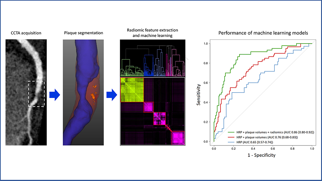
A total of 60 patients with acute MI prospectively underwent coronary CTA before invasive angiography and were matched to 60 patients with stable CAD. For all coronary lesions, high-risk plaque (HRP) characteristics were qualitatively assessed, followed by semiautomated plaque quantification and extraction of 1,103 radiomic features. Machine learning models were built to examine the additive value of radiomic features for discriminating culprit lesions over and above HRP and plaque volumes.

RESULTS

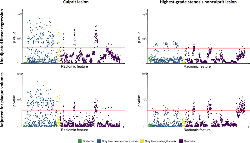
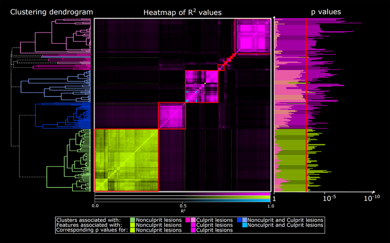
Culprit lesions had higher mean volumes of noncalcified plaque (NCP) and low-density noncalcified plaque (LDNCP) compared with the highest-grade stenosis nonculprits and highest-grade stenosis stable CAD lesions (NCP: 138.1 mm vs 110.7 mm vs 102.7 mm; LDNCP: 14.2 mm vs 9.8 mm vs 8.4 mm; both P < 0.01). In multivariable linear regression adjusted for NCP and LDNCP volumes, 14.9% (164 of 1,103) of radiomic features were associated with culprits and 9.7% (107 of 1,103) were associated with the highest-grade stenosis nonculprits (critical P < 0.0007) when compared with highest-grade stenosis stable CAD lesions as reference. Hierarchical clustering of significant radiomic features identified 9 unique data clusters (latent phenotypes): 5 contained radiomic features specific to culprits, 1 contained features specific to highest-grade stenosis nonculprits, and 3 contained features associated with either lesion type. Radiomic features provided incremental value for discriminating culprit lesions when added to a machine learning model containing HRP and plaque volumes (area under the receiver-operating characteristic curve 0.86 vs 0.76; P = 0.004).

CONCLUSIONS

Culprit lesions and highest-grade stenosis nonculprit lesions in MI have distinct radiomic signatures compared with lesions in stable CAD. Within the vulnerable patient may exist individual vulnerable plaques identifiable by coronary CTA-based precision phenotyping.

摘要

目的

本研究旨在通过基于冠状动脉计算机断层扫描血管造影(CTA)的放射组学分析,精确表型心肌梗死(MI)中的罪犯和非罪犯病变以及稳定型冠状动脉疾病(CAD)中的病变。

背景

目前仍存在争议,即易损患者体内的任何单个冠状动脉粥样硬化斑块是否具有独特的形态,从而增加临床事件的风险。

方法

前瞻性纳入 60 例急性 MI 患者,在进行有创血管造影前进行冠状动脉 CTA 检查,并与 60 例稳定型 CAD 患者相匹配。对所有冠状动脉病变进行高风险斑块(HRP)特征的定性评估,然后进行半自动斑块定量和提取 1103 个放射组学特征。构建机器学习模型,以检查放射组学特征在区分罪犯病变方面的附加价值,超过 HRP 和斑块体积。

结果

与最高狭窄度非罪犯病变和最高狭窄度稳定 CAD 病变相比,罪犯病变的非钙化斑块(NCP)和低密度非钙化斑块(LDNCP)平均体积更高(NCP:138.1mm 比 110.7mm 比 102.7mm;LDNCP:14.2mm 比 9.8mm 比 8.4mm;均 P<0.01)。在多变量线性回归中,校正 NCP 和 LDNCP 体积后,14.9%(1103 个中的 164 个)的放射组学特征与罪犯病变相关,9.7%(1103 个中的 107 个)与最高狭窄度非罪犯病变相关(临界 P<0.0007),以最高狭窄度稳定 CAD 病变作为参考。对显著放射组学特征进行层次聚类,可识别出 9 个独特的数据簇(潜在表型):5 个簇包含与罪犯病变特异性相关的放射组学特征,1 个簇包含与最高狭窄度非罪犯病变特异性相关的特征,3 个簇包含与两种病变类型均相关的特征。当放射组学特征添加到包含 HRP 和斑块体积的机器学习模型中时,可为区分罪犯病变提供附加价值(接受者操作特征曲线下面积 0.86 比 0.76;P=0.004)。

结论

MI 中的罪犯病变和最高狭窄度非罪犯病变与稳定 CAD 中的病变具有明显不同的放射组学特征。在易损患者中,可能存在可通过基于冠状动脉 CTA 的精准表型识别的个体易损斑块。

https://cdn.ncbi.nlm.nih.gov/pmc/blobs/ec9b/9072980/13f6e76cff9c/nihms-1768553-f0004.jpg
https://cdn.ncbi.nlm.nih.gov/pmc/blobs/ec9b/9072980/b2824b11a4fe/nihms-1768553-f0001.jpg
https://cdn.ncbi.nlm.nih.gov/pmc/blobs/ec9b/9072980/be43f5814f86/nihms-1768553-f0002.jpg
https://cdn.ncbi.nlm.nih.gov/pmc/blobs/ec9b/9072980/d477e34036f4/nihms-1768553-f0003.jpg
https://cdn.ncbi.nlm.nih.gov/pmc/blobs/ec9b/9072980/13f6e76cff9c/nihms-1768553-f0004.jpg

相似文献

1
Radiomics-Based Precision Phenotyping Identifies Unstable Coronary Plaques From Computed Tomography Angiography.基于放射组学的精准表型分析从计算机断层血管造影术识别不稳定的冠状动脉斑块。
JACC Cardiovasc Imaging. 2022 May;15(5):859-871. doi: 10.1016/j.jcmg.2021.11.016. Epub 2022 Jan 12.
2
Coronary Computed Tomography Angiography-Specific Definitions of High-Risk Plaque Features Improve Detection of Acute Coronary Syndrome.冠状动脉计算机断层扫描血管造影术特定的高危斑块特征定义可提高急性冠状动脉综合征的检出率。
Circ Cardiovasc Imaging. 2018 Aug;11(8):e007657. doi: 10.1161/CIRCIMAGING.118.007657.
3
Identification of noncalcified coronary plaque characteristics using machine learning radiomic analysis of non-contrast high-resolution computed tomography.使用非对比高分辨率计算机断层扫描的机器学习放射组学分析识别非钙化性冠状动脉斑块特征。
Kardiol Pol. 2023;81(10):978-989. doi: 10.33963/v.kp.97206. Epub 2023 Sep 3.
4
Identification of High-Risk Plaques Destined to Cause Acute Coronary Syndrome Using Coronary Computed Tomographic Angiography and Computational Fluid Dynamics.采用冠状动脉计算机断层扫描血管造影和计算流体力学识别易致急性冠状动脉综合征的高危斑块。
JACC Cardiovasc Imaging. 2019 Jun;12(6):1032-1043. doi: 10.1016/j.jcmg.2018.01.023. Epub 2018 Mar 14.
5
Plaque Morphology as Predictor of Late Plaque Events in Patients With Asymptomatic Type 2 Diabetes: A Long-Term Observational Study.斑块形态作为无症状 2 型糖尿病患者晚期斑块事件的预测因子:一项长期观察研究。
JACC Cardiovasc Imaging. 2019 Jul;12(7 Pt 2):1353-1363. doi: 10.1016/j.jcmg.2018.02.025. Epub 2018 May 16.
6
Pericoronary Adipose Tissue Computed Tomography Attenuation and High-Risk Plaque Characteristics in Acute Coronary Syndrome Compared With Stable Coronary Artery Disease.对比稳定型冠心病,急性冠状动脉综合征患者的冠状动脉旁脂肪组织 CT 衰减值与高危斑块特征。
JAMA Cardiol. 2018 Sep 1;3(9):858-863. doi: 10.1001/jamacardio.2018.1997.
7
CT-based total vessel plaque analyses improves prediction of hemodynamic significance lesions as assessed by fractional flow reserve in patients with stable angina pectoris.基于 CT 的全血管斑块分析可改善对稳定性心绞痛患者血流动力学意义病变的预测,这些病变通过血流储备分数进行评估。
J Cardiovasc Comput Tomogr. 2018 Jul-Aug;12(4):344-349. doi: 10.1016/j.jcct.2018.04.008. Epub 2018 May 8.
8
Myocardial Infarction Associates With a Distinct Pericoronary Adipose Tissue Radiomic Phenotype: A Prospective Case-Control Study.心肌梗死与独特的冠状动脉周围脂肪组织放射组学表型相关:一项前瞻性病例对照研究。
JACC Cardiovasc Imaging. 2020 Nov;13(11):2371-2383. doi: 10.1016/j.jcmg.2020.06.033. Epub 2020 Aug 26.
9
Comparison of quantitative atherosclerotic plaque burden from coronary CT angiography in patients with first acute coronary syndrome and stable coronary artery disease.首次急性冠状动脉综合征患者与稳定型冠状动脉疾病患者冠状动脉CT血管造影定量动脉粥样硬化斑块负荷的比较。
J Cardiovasc Comput Tomogr. 2014 Sep-Oct;8(5):368-74. doi: 10.1016/j.jcct.2014.07.007. Epub 2014 Aug 23.
10
Pericoronary Adipose Tissue Attenuation in Patients With Acute Coronary Syndrome Versus Stable Coronary Artery Disease.急性冠状动脉综合征与稳定型冠状动脉疾病患者的冠状动脉旁脂肪组织衰减。
Circ Cardiovasc Imaging. 2023 Feb;16(2):e014672. doi: 10.1161/CIRCIMAGING.122.014672. Epub 2023 Feb 3.

引用本文的文献

1
An artificial intelligence model integrating culprit lesion diagnosis and risk assessment for acute coronary syndrome.一种整合急性冠状动脉综合征罪犯病变诊断与风险评估的人工智能模型。
EClinicalMedicine. 2025 Aug 12;87:103415. doi: 10.1016/j.eclinm.2025.103415. eCollection 2025 Sep.
2
The Machine Learning Models in Major Cardiovascular Adverse Events Prediction Based on Coronary Computed Tomography Angiography: Systematic Review.基于冠状动脉计算机断层扫描血管造影术的主要心血管不良事件预测中的机器学习模型:系统评价
J Med Internet Res. 2025 Jun 13;27:e68872. doi: 10.2196/68872.
3
Artificial Intelligence in Coronary Artery Interventions: Preprocedural Planning and Procedural Assistance.

本文引用的文献

1
Contribution of Risk Factors to the Development of Coronary Atherosclerosis as Confirmed via Coronary CT Angiography: A Longitudinal Radiomics-based Study.基于冠脉 CT 血管造影的风险因素对冠状动脉粥样硬化发展的贡献:一项纵向基于放射组学的研究。
Radiology. 2021 Apr;299(1):97-106. doi: 10.1148/radiol.2021203179. Epub 2021 Feb 16.
2
Percutaneous Coronary Intervention for Vulnerable Coronary Atherosclerotic Plaque.经皮冠状动脉介入治疗易损冠状动脉粥样硬化斑块
J Am Coll Cardiol. 2020 Nov 17;76(20):2289-2301. doi: 10.1016/j.jacc.2020.09.547. Epub 2020 Oct 15.
3
Current Evidence and Recommendations for Coronary CTA First in Evaluation of Stable Coronary Artery Disease.
冠状动脉介入治疗中的人工智能:术前规划与术中辅助
J Soc Cardiovasc Angiogr Interv. 2025 Mar 18;4(3Part B):102519. doi: 10.1016/j.jscai.2024.102519. eCollection 2025 Mar.
4
Beyond plaque segmentation: a combined radiomics-deep learning approach for automated CAD-RADS classification.超越斑块分割:一种用于自动CAD-RADS分类的放射组学与深度学习相结合的方法。
Front Med (Lausanne). 2025 Mar 26;12:1536239. doi: 10.3389/fmed.2025.1536239. eCollection 2025.
5
Quantitative plaque characterization, pericoronary fat attenuation index, and fractional flow reserve: a novel method for differentiating between stable and unstable angina pectoris in a case-control study.定量斑块特征、冠状动脉周围脂肪衰减指数和血流储备分数:一项病例对照研究中区分稳定型和不稳定型心绞痛的新方法。
Quant Imaging Med Surg. 2025 Feb 1;15(2):1139-1150. doi: 10.21037/qims-24-1031. Epub 2025 Jan 22.
6
A Scoping Review of Machine-Learning Derived Radiomic Analysis of CT and PET Imaging to Investigate Atherosclerotic Cardiovascular Disease.基于 CT 和 PET 成像的机器学习衍生放射组学分析用于研究动脉粥样硬化性心血管疾病的范围综述。
Tomography. 2024 Sep 3;10(9):1455-1487. doi: 10.3390/tomography10090108.
7
Computed Tomography Angiography Identified High-Risk Coronary Plaques: From Diagnosis to Prognosis and Future Management.计算机断层扫描血管造影术识别出的高危冠状动脉斑块:从诊断到预后及未来管理
Diagnostics (Basel). 2024 Aug 1;14(15):1671. doi: 10.3390/diagnostics14151671.
8
Advances in Artificial Intelligence-Assisted Coronary Computed Tomographic Angiography for Atherosclerotic Plaque Characterization.人工智能辅助冠状动脉计算机断层扫描血管造影在动脉粥样硬化斑块特征分析方面的进展。
Rev Cardiovasc Med. 2024 Jan 15;25(1):27. doi: 10.31083/j.rcm2501027. eCollection 2024 Jan.
9
Application of Quantitative Assessment of Coronary Atherosclerosis by Coronary Computed Tomographic Angiography.冠状动脉 CT 血管造影定量评估冠状动脉粥样硬化的应用。
Korean J Radiol. 2024 Jun;25(6):518-539. doi: 10.3348/kjr.2023.1311.
10
Noninvasive Atherosclerotic Phenotyping: The Next Frontier into Understanding the Pathobiology of Coronary Artery Disease.无创性动脉粥样硬化表型分析:深入了解冠状动脉疾病病理生物学的下一个前沿领域。
Curr Atheroscler Rep. 2024 Jul;26(7):305-315. doi: 10.1007/s11883-024-01205-7. Epub 2024 May 10.
冠状动脉CTA首次用于稳定型冠状动脉疾病评估的当前证据与建议
J Am Coll Cardiol. 2020 Sep 15;76(11):1358-1362. doi: 10.1016/j.jacc.2020.06.078.
4
Nonculprit Lesion Severity and Outcome of Revascularization in Patients With STEMI and Multivessel Coronary Disease.非罪犯病变严重程度与 STEMI 及多支血管病变患者血运重建的结局。
J Am Coll Cardiol. 2020 Sep 15;76(11):1277-1286. doi: 10.1016/j.jacc.2020.07.034.
5
Myocardial Infarction Associates With a Distinct Pericoronary Adipose Tissue Radiomic Phenotype: A Prospective Case-Control Study.心肌梗死与独特的冠状动脉周围脂肪组织放射组学表型相关:一项前瞻性病例对照研究。
JACC Cardiovasc Imaging. 2020 Nov;13(11):2371-2383. doi: 10.1016/j.jcmg.2020.06.033. Epub 2020 Aug 26.
6
Nonculprit Lesion Plaque Morphology in Patients With ST-Segment-Elevation Myocardial Infarction: Results From the COMPLETE Trial Optical Coherence Tomography Substudys.ST段抬高型心肌梗死患者非罪犯病变的斑块形态:COMPLETE试验光学相干断层扫描子研究结果
Circ Cardiovasc Interv. 2020 Jul;13(7):e008768. doi: 10.1161/CIRCINTERVENTIONS.119.008768. Epub 2020 Jul 10.
7
Low-Attenuation Noncalcified Plaque on Coronary Computed Tomography Angiography Predicts Myocardial Infarction: Results From the Multicenter SCOT-HEART Trial (Scottish Computed Tomography of the HEART).冠状动脉计算机断层扫描血管造影中的低衰减非钙化斑块可预测心肌梗死:来自多中心 SCOT-HEART 试验(苏格兰心脏计算机断层扫描)的结果。
Circulation. 2020 May 5;141(18):1452-1462. doi: 10.1161/CIRCULATIONAHA.119.044720. Epub 2020 Mar 16.
8
From Detecting the Vulnerable Plaque to Managing the Vulnerable Patient: JACC State-of-the-Art Review.从检测易损斑块到管理易损患者:JACC 最新综述。
J Am Coll Cardiol. 2019 Sep 24;74(12):1582-1593. doi: 10.1016/j.jacc.2019.07.062.
9
Radiomics versus Visual and Histogram-based Assessment to Identify Atheromatous Lesions at Coronary CT Angiography: An ex Vivo Study.基于放射组学与视觉、直方图评估识别冠状动脉 CT 血管造影中的粥样硬化病变:一项离体研究。
Radiology. 2019 Oct;293(1):89-96. doi: 10.1148/radiol.2019190407. Epub 2019 Aug 6.
10
Standardized volumetric plaque quantification and characterization from coronary CT angiography: a head-to-head comparison with invasive intravascular ultrasound.从冠状动脉 CT 血管造影中进行标准化容积斑块定量和特征描述:与有创血管内超声的头对头比较。
Eur Radiol. 2019 Nov;29(11):6129-6139. doi: 10.1007/s00330-019-06219-3. Epub 2019 Apr 26.