Henderson Edward G A, Vasquez Osorio Eliana M, van Herk Marcel, Green Andrew F
The University of Manchester, Oxford Rd, Manchester M13 9PL, UK.
Radiotherapy Related Research, The Christie NHS Foundation Trust, Manchester M20 4BX, UK.
Phys Imaging Radiat Oncol. 2022 Apr 28;22:44-50. doi: 10.1016/j.phro.2022.04.003. eCollection 2022 Apr.
Convolutional neural networks (CNNs) are increasingly used to automate segmentation for radiotherapy planning, where accurate segmentation of organs-at-risk (OARs) is crucial. Training CNNs often requires large amounts of data. However, large, high quality datasets are scarce. The aim of this study was to develop a CNN capable of accurate head and neck (HN) 3D auto-segmentation of planning CT scans using a small training dataset (34 CTs).
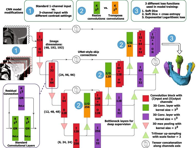
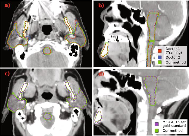
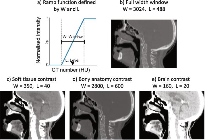
Elements of our custom CNN architecture were varied to optimise segmentation performance. We tested and evaluated the impact of: using multiple contrast channels for the CT scan input at specific soft tissue and bony anatomy windows, resize vs. transpose convolutions, and loss functions based on overlap metrics and cross-entropy in different combinations. Model segmentation performance was compared with the inter-observer deviation of two doctors' gold standard segmentations using the 95th percentile Hausdorff distance and mean distance-to-agreement (mDTA). The best performing configuration was further validated on a popular public dataset to compare with state-of-the-art (SOTA) auto-segmentation methods.
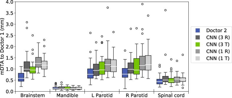
Our best performing CNN configuration was competitive with current SOTA methods when evaluated on the public dataset with mDTA of mm for the brainstem, mm for the mandible, mm for the left parotid and mm for the right parotid.
Through careful tuning and customisation we trained a 3D CNN with a small dataset to produce segmentations of HN OARs with an accuracy that is comparable with inter-clinician deviations. Our proposed model performed competitively with current SOTA methods.
卷积神经网络(CNN)越来越多地用于放疗计划的自动分割,其中危及器官(OAR)的准确分割至关重要。训练CNN通常需要大量数据。然而,大型高质量数据集稀缺。本研究的目的是开发一种能够使用小训练数据集(34例CT)对头部和颈部(HN)计划CT扫描进行准确三维自动分割的CNN。
对我们定制的CNN架构元素进行了多种变化以优化分割性能。我们测试并评估了以下因素的影响:在特定软组织和骨解剖窗口使用多个对比通道作为CT扫描输入、调整大小卷积与转置卷积,以及基于不同组合的重叠度量和交叉熵的损失函数。使用第95百分位数豪斯多夫距离和平均一致距离(mDTA)将模型分割性能与两位医生的金标准分割的观察者间偏差进行比较。在一个流行的公共数据集上进一步验证表现最佳的配置,以与当前的最先进(SOTA)自动分割方法进行比较。
当在公共数据集上进行评估时,我们表现最佳的CNN配置与当前SOTA方法具有竞争力,脑干的mDTA为 毫米,下颌骨为 毫米,左腮腺为 毫米,右腮腺为 毫米。
通过仔细调整和定制,我们使用小数据集训练了一个三维CNN,以产生与临床医生间偏差相当的HN OAR分割精度。我们提出的模型与当前SOTA方法具有竞争力。