Sharma Satyavan, Malavia Gunjan Arvindbhai
Department of Cardiology, Bombay Hospital and Medical Research Centre, Mumbai, Maharashtra, India.
Ann Pediatr Cardiol. 2021 Oct-Dec;14(4):496-500. doi: 10.4103/apc.apc_14_21. Epub 2022 Mar 25.
Infective endocarditis (IE) involving the native pulmonary valve (PV) is extremely rare, with no data in Indian literature. The objective of this communication is to describe the clinical and diagnostic characteristics, underlying risk factors, microbiological features, and management of PVIE.
This is a retrospective analysis of 8 cases of PVIE managed in a tertiary care center from 1992 to 2020.
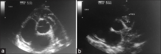
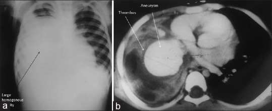
PVIE was observed in 8 patients with underlying congenital cardiac malformation (Group A, 6 Patients) and in patients with central venous catheter (Group B, 2 patients). All the patients had prolonged febrile illness accompanied by right heart failure 4 (50%), septic pulmonary emboli 2 (25%), and pulmonary regurgitation 3 (37.5%). Trans-thoracic echocardiography demonstrated the vegetations, whereas computed tomography of chest diagnosed pulmonary emboli in 2 (25%), and pulmonary artery aneurysm in 1 (12.5%) patient. The early mortality was extremely high (5, 62.5%). Delayed diagnosis, fulminant septicemia, and multi-organ failure resulted in unfavorable outcomes.
IE of the native PV is a rare and potentially lethal illness. Diagnosis should be considered in any febrile patient with an underlying congenital defect, central venous line, bacteremia, and comorbidities. Multi-modality imaging should be utilized to enhance the diagnostic yield and detect complications promptly.
累及原生肺动脉瓣(PV)的感染性心内膜炎(IE)极为罕见,印度文献中尚无相关数据。本文旨在描述原生肺动脉瓣感染性心内膜炎(PVIE)的临床和诊断特征、潜在危险因素、微生物学特征及治疗方法。
这是一项对1992年至2020年在一家三级医疗中心接受治疗的8例PVIE患者的回顾性分析。
8例PVIE患者中,6例(A组)有先天性心脏畸形,2例(B组)有中心静脉导管。所有患者均有长期发热性疾病,并伴有右心衰竭4例(50%)、脓毒性肺栓塞2例(25%)和肺动脉反流3例(37.5%)。经胸超声心动图显示有赘生物,胸部计算机断层扫描诊断出2例(25%)有肺栓塞,1例(12.5%)有肺动脉瘤。早期死亡率极高(5例,62.5%)。诊断延迟、暴发性败血症和多器官功能衰竭导致了不良后果。
原生肺动脉瓣IE是一种罕见且可能致命的疾病。对于任何有先天性缺陷、中心静脉导管、菌血症和合并症的发热患者,都应考虑进行诊断。应采用多模态成像来提高诊断率并及时发现并发症。