Chen Wei-Ming, Fu Min, Zhang Cheng-Ju, Xing Qing-Qing, Zhou Fei, Lin Meng-Jie, Dong Xuan, Huang Jiaofeng, Lin Su, Hong Mei-Zhu, Zheng Qi-Zhong, Pan Jin-Shui
Liver Research Center, The First Affiliated Hospital of Fujian Medical University, Fuzhou, China.
School of Medicine, Xiamen University, Xiamen, China.
Front Med (Lausanne). 2022 Apr 22;9:853261. doi: 10.3389/fmed.2022.853261. eCollection 2022.
We aim to develop a diagnostic tool for pathological-image classification using transfer learning that can be applied to diverse tumor types.
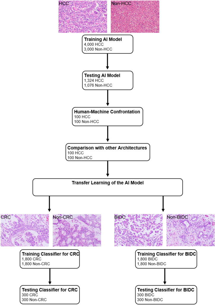
Microscopic images of liver tissue with and without hepatocellular carcinoma (HCC) were used to train and validate the classification framework based on a convolutional neural network. To evaluate the universal classification performance of the artificial intelligence (AI) framework, histological images from colorectal tissue and the breast were collected. Images for the training and validation sets were obtained from the Xiamen Hospital of Traditional Chinese Medicine, and those for the test set were collected from Zhongshan Hospital Xiamen University. The accuracy, sensitivity, and specificity values for the proposed framework were reported and compared with those of human image interpretation.
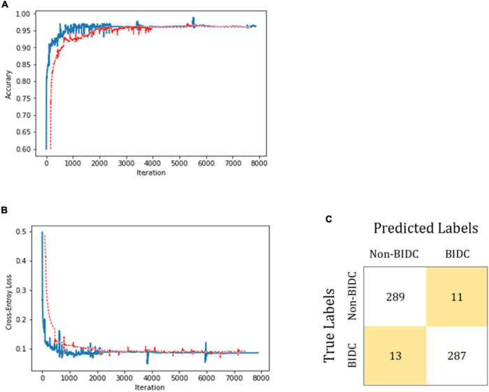
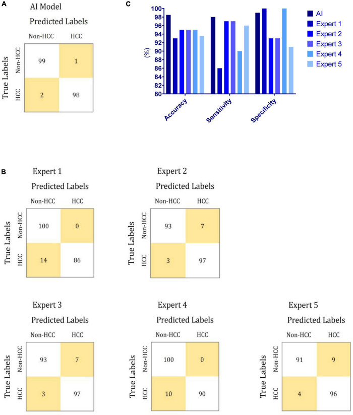
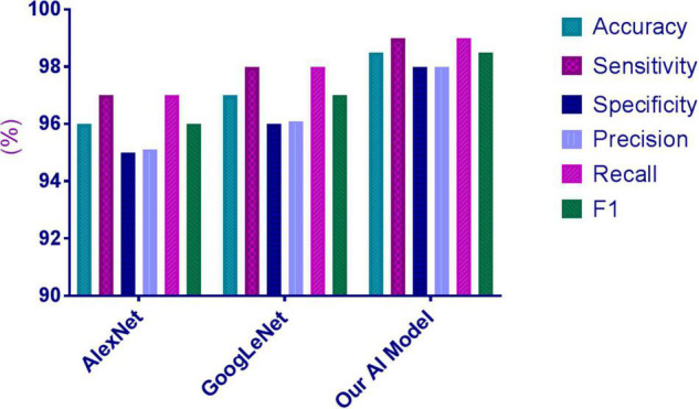
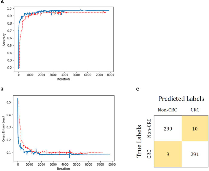
In the human-machine comparisons, the sensitivity, and specificity for the AI algorithm were 98.0, and 99.0%, whereas for the human experts, the sensitivity ranged between 86.0 and 97.0%, while the specificity ranged between 91.0 and 100%. Based on transfer learning, the accuracies of the AI framework in classifying colorectal carcinoma and breast invasive ductal carcinoma were 96.8 and 96.0%, respectively.
The performance of the proposed AI framework in classifying histological images with HCC was comparable to the classification performance achieved by human experts, indicating that extending the proposed AI's application to diagnoses and treatment recommendations is a promising area for future investigation.
我们旨在开发一种利用迁移学习的病理图像分类诊断工具,该工具可应用于多种肿瘤类型。
使用有无肝细胞癌(HCC)的肝组织显微图像来训练和验证基于卷积神经网络的分类框架。为了评估人工智能(AI)框架的通用分类性能,收集了结直肠组织和乳腺的组织学图像。训练集和验证集的图像取自厦门市中医院,测试集的图像则收集自厦门大学附属中山医院。报告了所提出框架的准确性、敏感性和特异性值,并与人工图像解读的结果进行比较。
在人机比较中,AI算法的敏感性和特异性分别为98.0%和99.0%,而人类专家的敏感性在86.0%至97.0%之间,特异性在91.0%至100%之间。基于迁移学习,AI框架对结直肠癌和乳腺浸润性导管癌的分类准确率分别为96.8%和96.0%。
所提出的AI框架在对伴有HCC的组织学图像进行分类方面的性能与人类专家的分类性能相当,这表明将所提出的AI应用扩展到诊断和治疗建议是未来一个有前景的研究领域。