Department of Computer Science, Stony Brook University, Stony Brook, NY, 11794, USA.
Department of Mathematics and Statistics, Georgia State University, Atlanta, GA, 30303, USA.
Sci Rep. 2021 Jan 8;11(1):139. doi: 10.1038/s41598-020-80610-9.
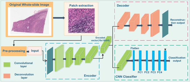
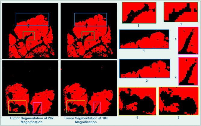
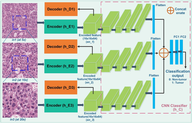
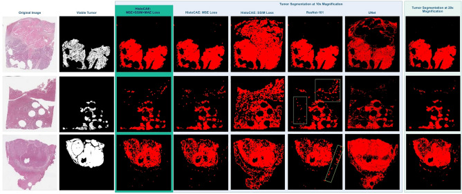
Liver cancer is one of the leading causes of cancer deaths in Asia and Africa. It is caused by the Hepatocellular carcinoma (HCC) in almost 90% of all cases. HCC is a malignant tumor and the most common histological type of the primary liver cancers. The detection and evaluation of viable tumor regions in HCC present an important clinical significance since it is a key step to assess response of chemoradiotherapy and tumor cell proportion in genetic tests. Recent advances in computer vision, digital pathology and microscopy imaging enable automatic histopathology image analysis for cancer diagnosis. In this paper, we present a multi-resolution deep learning model HistoCAE for viable tumor segmentation in whole-slide liver histopathology images. We propose convolutional autoencoder (CAE) based framework with a customized reconstruction loss function for image reconstruction, followed by a classification module to classify each image patch as tumor versus non-tumor. The resulting patch-based prediction results are spatially combined to generate the final segmentation result for each WSI. Additionally, the spatially organized encoded feature map derived from small image patches is used to compress the gigapixel whole-slide images. Our proposed model presents superior performance to other benchmark models with extensive experiments, suggesting its efficacy for viable tumor area segmentation with liver whole-slide images.
肝癌是亚洲和非洲癌症死亡的主要原因之一。几乎所有病例中,有 90%是由肝细胞癌 (HCC) 引起的。HCC 是一种恶性肿瘤,也是原发性肝癌最常见的组织学类型。检测和评估 HCC 中的存活肿瘤区域具有重要的临床意义,因为这是评估化学放射治疗反应和遗传测试中肿瘤细胞比例的关键步骤。计算机视觉、数字病理学和显微镜成像的最新进展使得癌症诊断的自动组织病理学图像分析成为可能。在本文中,我们提出了一种用于全切片肝组织病理学图像中存活肿瘤分割的多分辨率深度学习模型 HistoCAE。我们提出了一种基于卷积自动编码器 (CAE) 的框架,该框架具有用于图像重建的定制重建损失函数,随后是一个分类模块,用于将每个图像补丁分类为肿瘤与非肿瘤。基于补丁的预测结果在空间上进行组合,为每个 WSI 生成最终的分割结果。此外,从小图像补丁中提取的空间组织编码特征图用于压缩千兆像素的全切片图像。通过广泛的实验,我们提出的模型优于其他基准模型,这表明它在使用肝全切片图像进行存活肿瘤区域分割方面具有很好的效果。