Ageeli Wael, Zhang Xinyu, Ogbonnaya Chidozie N, Bray Susan E, Kernohan Neil M, Wilson Jennifer, Li Chunhui, Nabi Ghulam
Division of Imaging Sciences and Technology, School of Medicine, University of Dundee, Ninewells Hospital, Dundee, United Kingdom.
Diagnostic Radiology Department, College of Applied Medical Sciences, Jazan University, Jazan, Saudi Arabia.
Front Oncol. 2022 Apr 21;12:822476. doi: 10.3389/fonc.2022.822476. eCollection 2022.
Growing evidence suggests that the tumor microenvironment (TME) represented by cellular and acellular components plays a key role in the multistep process of metastases and response to therapies. However, imaging and molecular characterization of the TME in prostate cancer (PCa) and its role in predicting aggressive tumor behavior and disease progression is largely unexplored. The study explores the PCa TME through the characterization of cancer-associated fibroblasts (CAFs) using both immunohistochemistry (IHC) and genomics approaches. This is then correlated with transrectal ultrasound shear wave elastography (USWE)-measured tissue stiffness.
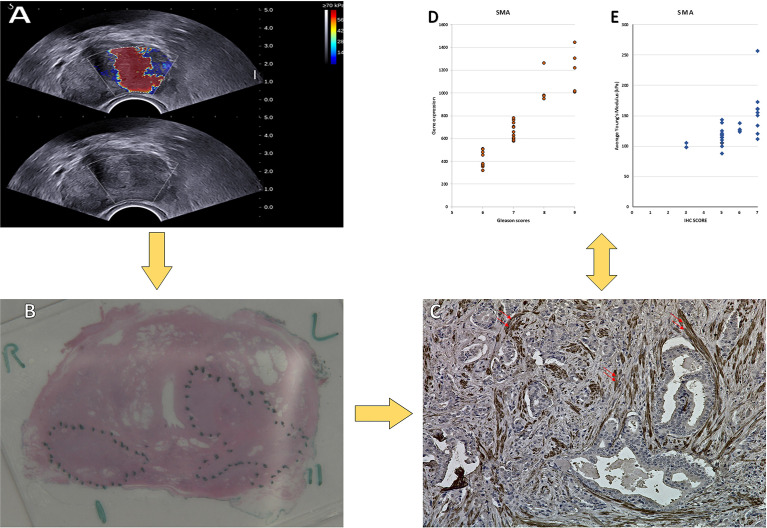
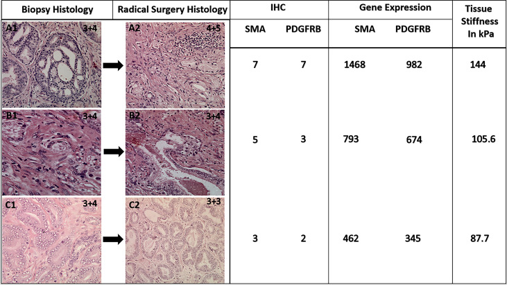
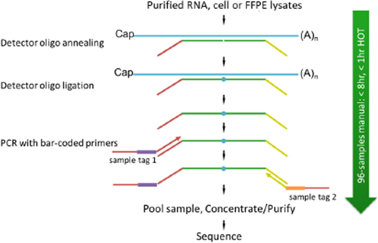
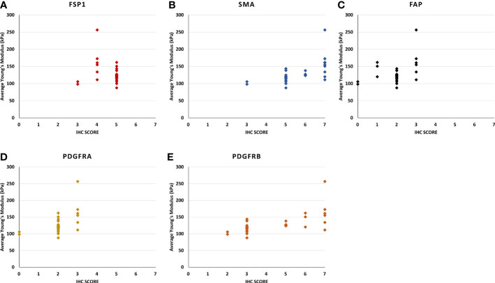
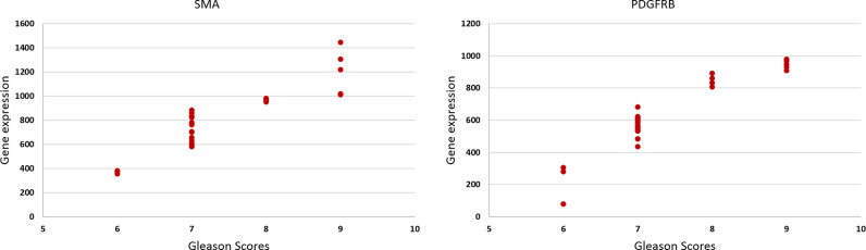
Thirty patients with clinically localized PCa undergoing radical prostatectomy for different risk categories of tumor (low, intermediate, and high) defined by Gleason score (GS) were prospectively recruited into this study. Prostatic tissue stiffness was measured using USWE prior to surgery. The CAFs within the TME were identified by IHC using a panel of six antibodies (FAP, SMAα, FSP1, CD36, PDGFRα, and PDGFRβ) as well as gene expression profiling using TempO-sequence analysis. Whether the pattern and degree of immunohistochemical positivity (measured by Quick score method) and expression of genes characterizing CAFs were correlated with USWE- and GS-measured tissue stiffnesses were tested using Spearman's rank correlation and Pearson correlation.
There was a statistically significant correlation between GS of cancers, the pattern of staining for CAFs by immunohistochemical staining, and tissue stiffness measured in kPa using USWE ( < 0.001). Significant differences were also observed in immunohistochemical staining patterns between normal prostate and prostatic cancerous tissue. PDGFRβ and SMAα immunostaining scores increased linearly with increasing the USWE stiffness and the GS of PCa. There was a significant positive correlation between increasing tissue stiffness in tumor stroma and SMAα and PDGFRβ gene expression in the fibromuscular stroma ( < 0.001).
USWE-measured tissue stiffness correlates with increased SMAα and PDGFRβ expressing CAFs and PCa GSs. This mechanistic correlation could be used for predicting the upgrading of GS from biopsies to radical surgery and response to novel treatments.
越来越多的证据表明,由细胞和无细胞成分构成的肿瘤微环境(TME)在转移的多步骤过程及对治疗的反应中起着关键作用。然而,前列腺癌(PCa)中TME的成像和分子特征及其在预测侵袭性肿瘤行为和疾病进展中的作用在很大程度上尚未得到探索。本研究通过使用免疫组织化学(IHC)和基因组学方法对癌症相关成纤维细胞(CAF)进行特征分析,来探究PCa的TME。然后将其与经直肠超声剪切波弹性成像(USWE)测量的组织硬度相关联。
前瞻性招募了30例因不同风险类别的肿瘤(根据Gleason评分(GS)定义为低、中、高风险)而接受根治性前列腺切除术的临床局限性PCa患者进入本研究。术前使用USWE测量前列腺组织硬度。通过使用一组六种抗体(FAP、SMAα、FSP1、CD36、PDGFRα和PDGFRβ)的IHC以及使用TempO-序列分析的基因表达谱来鉴定TME内的CAF。使用Spearman等级相关性和Pearson相关性检验免疫组织化学阳性的模式和程度(通过快速评分法测量)以及表征CAF的基因表达是否与USWE和GS测量的组织硬度相关。
癌症的GS、CAF免疫组织化学染色模式与使用USWE以kPa为单位测量的组织硬度之间存在统计学上的显著相关性(<0.001)。在正常前列腺组织和前列腺癌组织之间的免疫组织化学染色模式中也观察到显著差异。PDGFRβ和SMAα免疫染色评分随着USWE硬度和PCa的GS增加而线性增加。肿瘤基质中组织硬度增加与纤维肌基质中SMAα和PDGFRβ基因表达之间存在显著正相关(<0.001)。
USWE测量的组织硬度与表达SMAα和PDGFRβ的CAF增加以及PCa的GS相关。这种机制相关性可用于预测GS从活检到根治性手术的升级以及对新治疗的反应。