Department of Paediatric Orthopaedic; Centre for Stem Cell Research, Christian Medical College, Vellore, Tamil Nadu, India.
Indian J Med Res. 2021 May;154(5):732-742. doi: 10.4103/ijmr.IJMR_93_19.
BACKGROUND & OBJECTIVES: Rabbit model is commonly used to demonstrate the proof of concept in cartilage tissue engineering. However, limited studies have attempted to find an ideal source of rabbit mesenchymal stem cells (MSCs) for cartilage repair. This study aimed to compare the in vitro chondrogenic potential of rabbit MSCs isolated from three sources namely infrapatellar fat pad (IFP), periosteum (P) and bone marrow (BM).
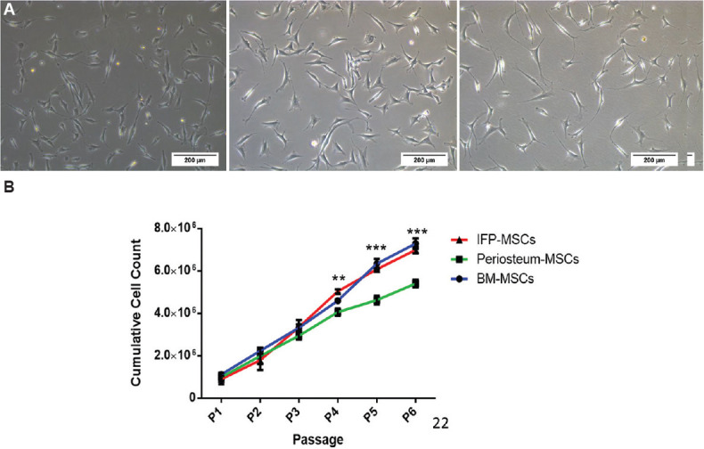
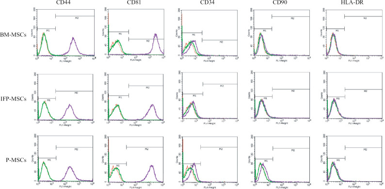
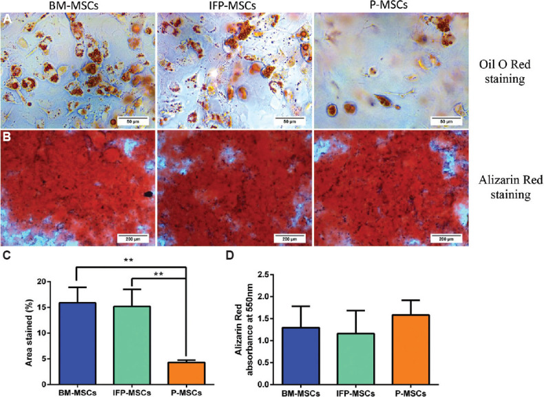
Rabbit MSCs from three sources were isolated and characterized using flow cytometry and multi-lineage differentiation assay. Cell proliferation was assessed using trypan blue dye exclusion test; in vitro chondrogenic potential was evaluated by histology and gene expression and the outcomes were compared amongst the three MSC sources.
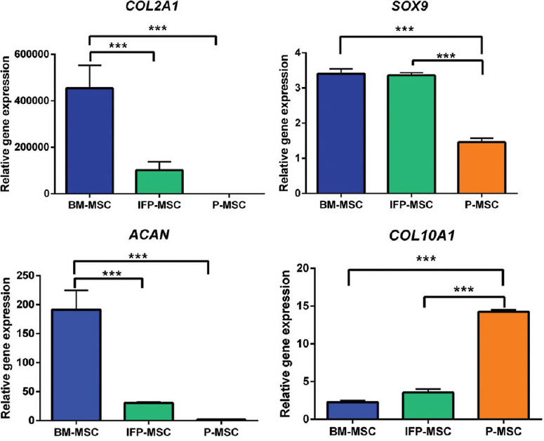
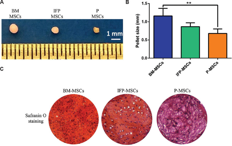
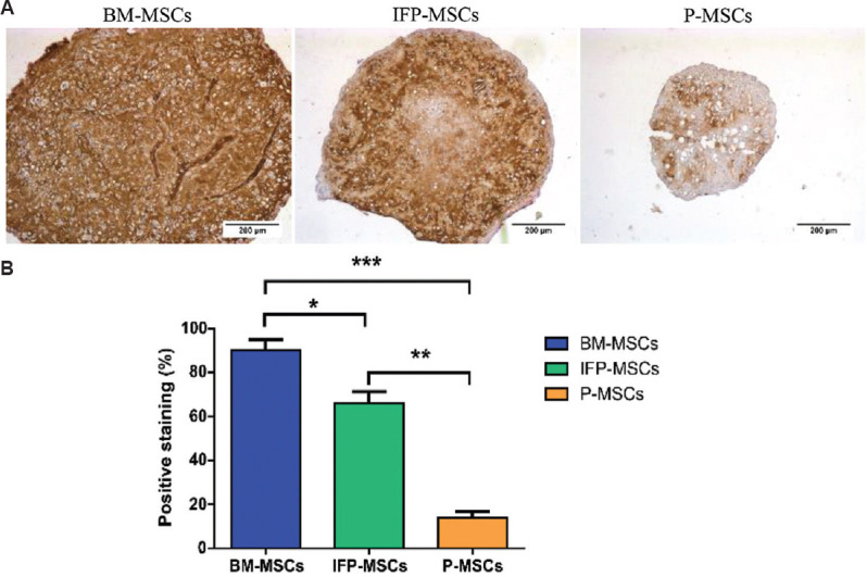
MSCs from three sources shared similar morphology and expressed >99 per cent positive for CD44 and CD81 and <3 per cent positive for negative markers CD34, CD90 and human leukocyte antigen - DR isotype (HLA-DR). The BM-MSCs and IFP-MSCs showed significantly higher cell proliferation (P<0.001) than the P-MSCs from passage 4. Histologically, BM-MSCs formed a thicker cartilage pellet (P<0.01) with abundant matrix deposition than IFP and P-MSCs during chondrogenic differentiation. The collagen type 2 staining was significantly (P<0.05) higher in BM-MSCs than the other two sources. These outcomes were further confirmed by gene expression, where the BM-MSCs demonstrated significantly higher expression (P<0.01) of cartilage-specific markers (COL2A1, SOX9 and ACAN) with less hypertrophy.
INTERPRETATION & CONCLUSIONS: This study demonstrated that BM-MSCs had superior chondrogenic potential and generated better cartilage than IFP and P-MSCs in rabbits. Thus, BM-MSCs remain a promising candidate for rabbit articular cartilage regeneration.
兔模型常用于证明软骨组织工程的概念验证。然而,很少有研究试图寻找用于软骨修复的理想兔间充质干细胞(MSCs)来源。本研究旨在比较三种来源(髌下脂肪垫(IFP)、骨膜(P)和骨髓(BM))分离的兔 MSCs 的体外成软骨潜力。
使用流式细胞术和多系分化试验分离和鉴定三种来源的兔 MSCs。使用台盼蓝染料排除试验评估细胞增殖;通过组织学和基因表达评估体外成软骨潜力,并比较三种 MSC 来源的结果。
三种来源的 MSCs 具有相似的形态,并且 >99%表达 CD44 和 CD81,<3%表达阴性标志物 CD34、CD90 和人类白细胞抗原-DR 同种型(HLA-DR)。从第 4 代开始,BM-MSCs 和 IFP-MSCs 的细胞增殖率显著高于 P-MSCs(P<0.001)。组织学上,BM-MSCs 在软骨分化过程中形成更厚的软骨球(P<0.01),且基质沉积丰富。BM-MSCs 的胶原 2 染色明显高于其他两种来源(P<0.05)。这些结果通过基因表达进一步证实,其中 BM-MSCs 表现出明显更高的软骨特异性标志物(COL2A1、SOX9 和 ACAN)表达(P<0.01),且较少发生肥大。
本研究表明,BM-MSCs 具有更强的成软骨潜力,在兔中比 IFP 和 P-MSCs 生成更好的软骨。因此,BM-MSCs 仍然是兔关节软骨再生的有前途的候选物。