• 文献检索
  • 文档翻译
  • 深度研究
  • 学术资讯
  • Suppr Zotero 插件Zotero 插件
  • 邀请有礼
  • 套餐&价格
  • 历史记录
应用&插件
Suppr Zotero 插件Zotero 插件浏览器插件Mac 客户端Windows 客户端微信小程序
定价
高级版会员购买积分包购买API积分包
服务
文献检索文档翻译深度研究API 文档MCP 服务
关于我们
关于 Suppr公司介绍联系我们用户协议隐私条款
关注我们

Suppr 超能文献

核心技术专利:CN118964589B侵权必究
粤ICP备2023148730 号-1Suppr @ 2026

文献检索

告别复杂PubMed语法,用中文像聊天一样搜索,搜遍4000万医学文献。AI智能推荐,让科研检索更轻松。

立即免费搜索

文件翻译

保留排版,准确专业,支持PDF/Word/PPT等文件格式,支持 12+语言互译。

免费翻译文档

深度研究

AI帮你快速写综述,25分钟生成高质量综述,智能提取关键信息,辅助科研写作。

立即免费体验

氧化铜纳米颗粒通过错误折叠铜锌超氧化物歧化酶 1(SOD1)引发巨噬细胞死亡。

Copper oxide nanoparticles trigger macrophage cell death with misfolding of Cu/Zn superoxide dismutase 1 (SOD1).

机构信息

Division of Molecular Toxicology, Institute of Environmental Medicine, Karolinska Institutet, Nobels väg 13, Stockholm, Sweden.

Department of Biotechnology and Life Sciences, University of Insubria, Varese, Italy.

出版信息

Part Fibre Toxicol. 2022 May 10;19(1):33. doi: 10.1186/s12989-022-00467-w.

DOI:10.1186/s12989-022-00467-w
PMID:35538581
原文链接:https://pmc.ncbi.nlm.nih.gov/articles/PMC9088059/
Abstract

BACKGROUND

Copper oxide (CuO) nanoparticles (NPs) are known to trigger cytotoxicity in a variety of cell models, but the mechanism of cell death remains unknown. Here we addressed the mechanism of cytotoxicity in macrophages exposed to CuO NPs versus copper chloride (CuCl).

METHODS

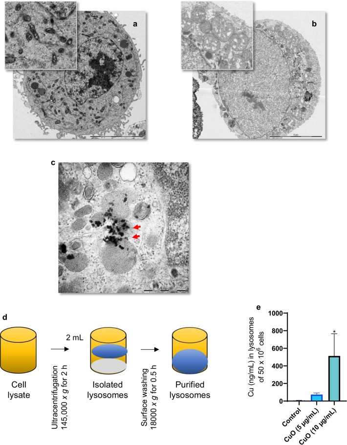
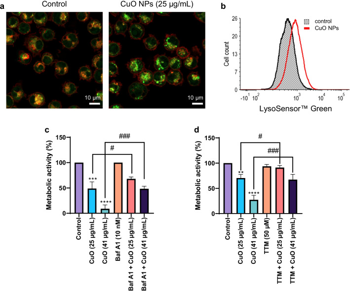
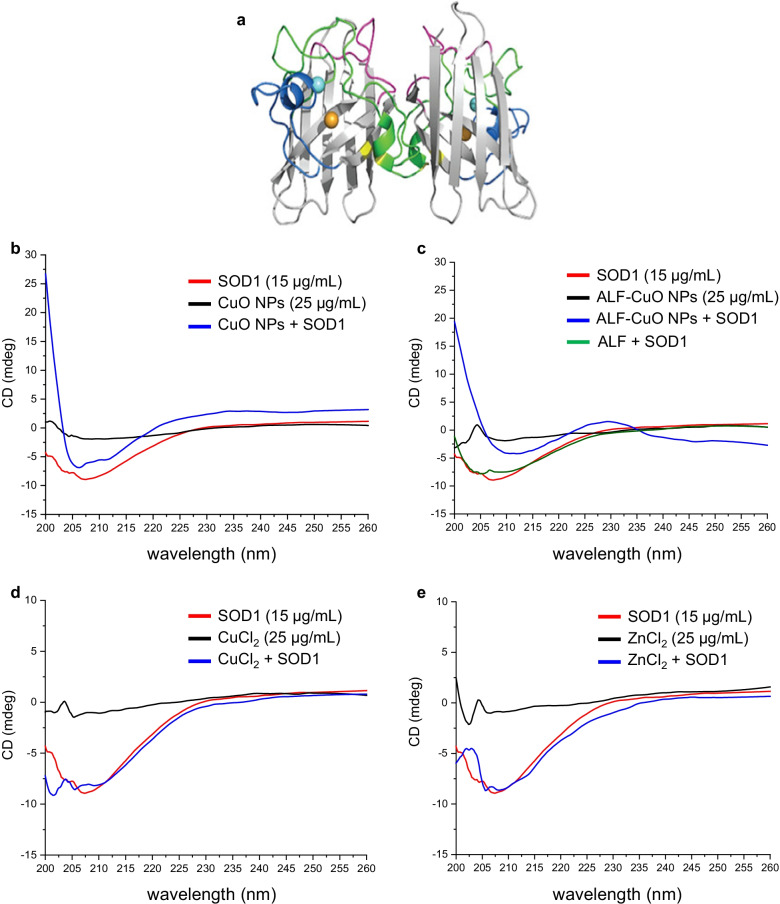
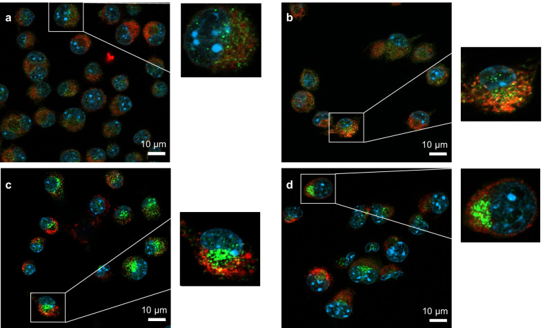
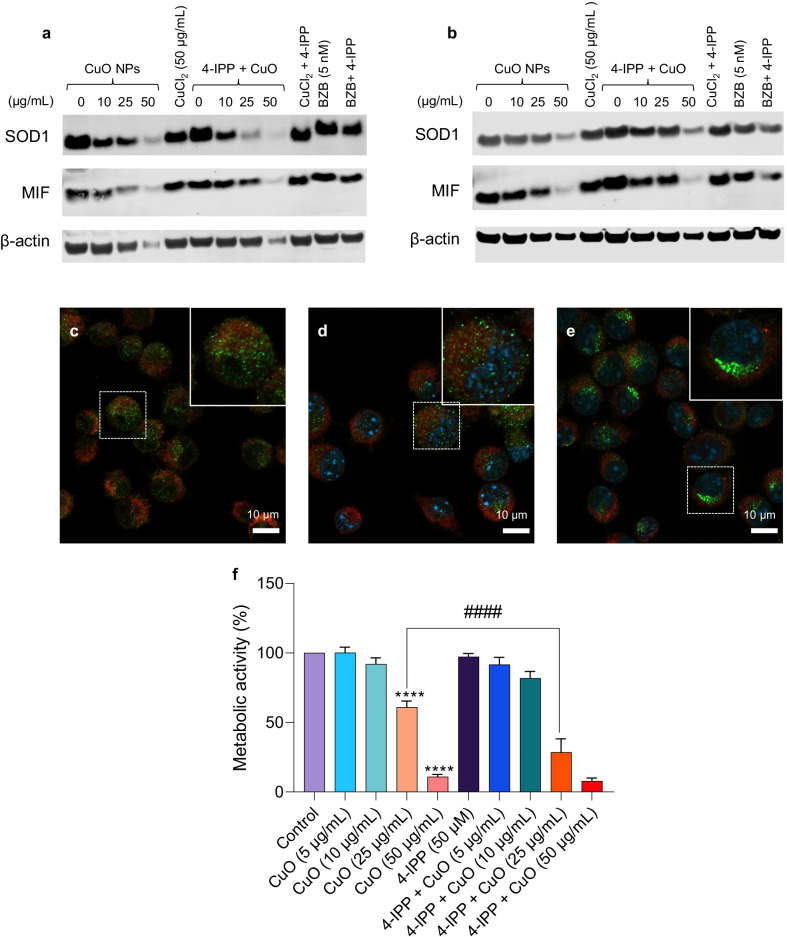
The mouse macrophage cell line RAW264.7 was used as an in vitro model. Particle uptake and the cellular dose of Cu were investigated by transmission electron microscopy (TEM) and inductively coupled plasma mass spectrometry (ICP-MS), respectively. The deposition of Cu in lysosomes isolated from macrophages was also determined by ICP-MS. Cell viability (metabolic activity) was assessed using the Alamar Blue assay, and oxidative stress was monitored by a variety of methods including a luminescence-based assay for cellular glutathione (GSH), and flow cytometry-based detection of mitochondrial superoxide and mitochondrial membrane potential. Protein aggregation was determined by confocal microscopy using an aggresome-specific dye and protein misfolding was determined by circular dichroism (CD) spectroscopy. Lastly, proteasome activity was investigated using a fluorometric assay.

RESULTS

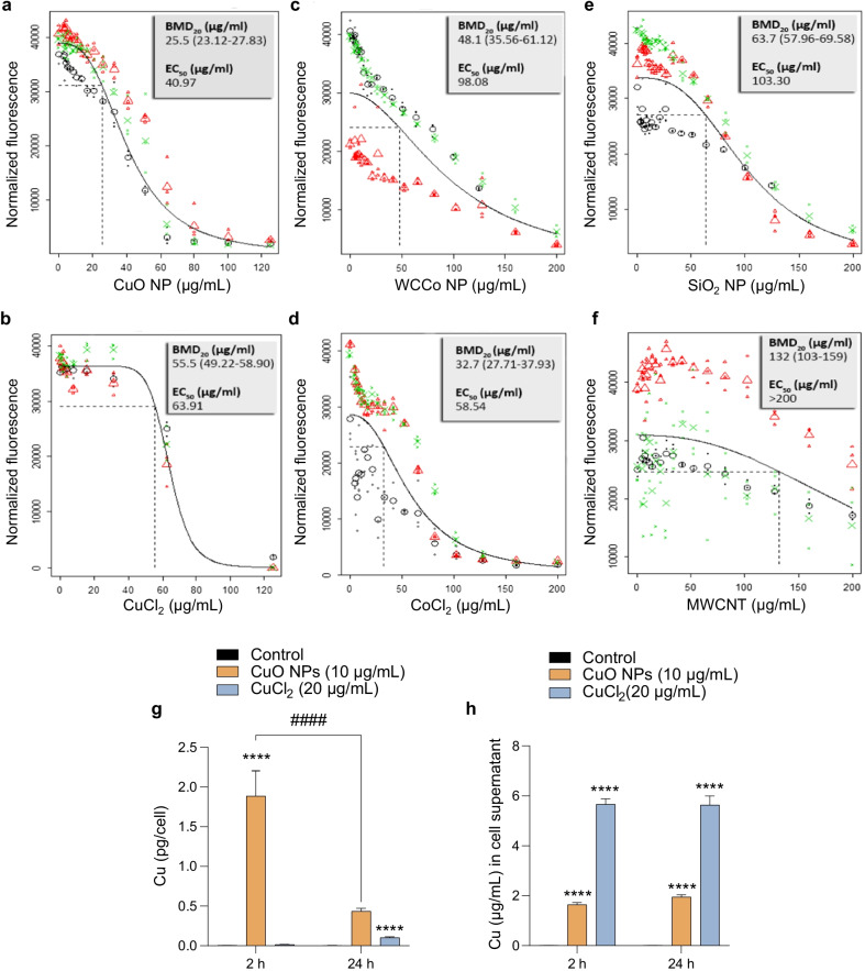
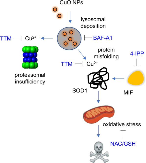
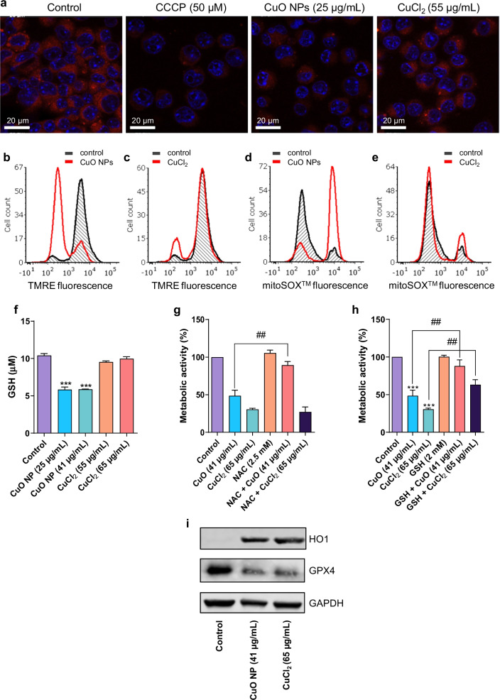
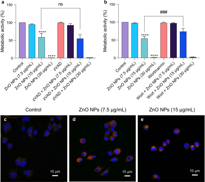
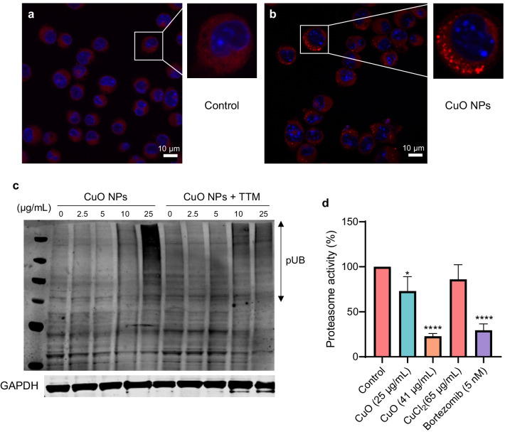
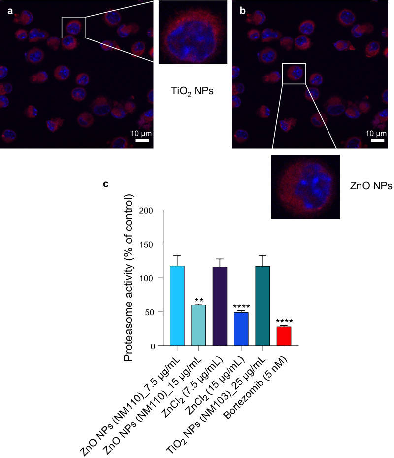
We observed rapid cellular uptake of CuO NPs in macrophages with deposition in lysosomes. CuO NP-elicited cell death was characterized by mitochondrial swelling with signs of oxidative stress including the production of mitochondrial superoxide and cellular depletion of GSH. We also observed a dose-dependent accumulation of polyubiquitinated proteins and loss of proteasomal function in CuO NP-exposed cells, and we could demonstrate misfolding and mitochondrial translocation of superoxide dismutase 1 (SOD1), a Cu/Zn-dependent enzyme that plays a pivotal role in the defense against oxidative stress. The chelation of copper ions using tetrathiomolybdate (TTM) prevented cell death whereas inhibition of the cellular SOD1 chaperone aggravated toxicity. Moreover, CuO NP-triggered cell death was insensitive to the pan-caspase inhibitor, zVAD-fmk, and to wortmannin, an inhibitor of autophagy, implying that this was a non-apoptotic cell death. ZnO NPs, on the other hand, triggered autophagic cell death.

CONCLUSIONS

CuO NPs undergo dissolution in lysosomes leading to copper-dependent macrophage cell death characterized by protein misfolding and proteasomal insufficiency. Specifically, we present novel evidence for Cu-induced SOD1 misfolding which accords with the pronounced oxidative stress observed in CuO NP-exposed macrophages. These results are relevant for our understanding of the consequences of inadvertent human exposure to CuO NPs.

摘要

背景

氧化铜 (CuO) 纳米颗粒 (NPs) 已被证实可在多种细胞模型中引发细胞毒性,但细胞死亡的机制仍不清楚。在此,我们研究了巨噬细胞暴露于 CuO NPs 与氯化铜 (CuCl) 时的细胞毒性机制。

方法

采用小鼠巨噬细胞系 RAW264.7 作为体外模型。通过透射电子显微镜 (TEM) 分别研究了颗粒摄取和细胞内铜含量,通过电感耦合等离子体质谱法 (ICP-MS) 研究了从巨噬细胞中分离的溶酶体中铜的沉积。还通过 ICP-MS 确定了铜在溶酶体中的沉积。通过阿尔玛蓝 (Alamar Blue) 测定法评估细胞活力(代谢活性),通过多种方法监测氧化应激,包括基于发光的细胞内谷胱甘肽 (GSH) 测定法和基于流式细胞术的线粒体超氧化物和线粒体膜电位检测。使用含有聚集物特异性染料的共聚焦显微镜确定蛋白聚集,使用圆二色性 (CD) 光谱法确定蛋白错误折叠。最后,使用荧光测定法研究蛋白酶体活性。

结果

我们观察到巨噬细胞中 CuO NPs 的快速细胞摄取,并沉积在溶酶体中。CuO NP 诱导的细胞死亡的特征是线粒体肿胀,伴有氧化应激的迹象,包括线粒体超氧化物的产生和细胞内 GSH 的消耗。我们还观察到 CuO NP 暴露细胞中多泛素化蛋白的剂量依赖性积累和蛋白酶体功能丧失,并且能够证明超氧化物歧化酶 1 (SOD1) 的错误折叠和线粒体易位,SOD1 是一种铜/锌依赖性酶,在抵御氧化应激中起关键作用。使用四硫钼酸盐 (TTM) 螯合铜离子可防止细胞死亡,而细胞 SOD1 伴侣抑制剂加重了毒性。此外,CuO NP 触发的细胞死亡对泛半胱天冬酶抑制剂 zVAD-fmk 不敏感,对自噬抑制剂wortmannin 不敏感,这表明这是一种非凋亡性细胞死亡。另一方面,氧化锌 (ZnO) NPs 引发自噬性细胞死亡。

结论

CuO NPs 在溶酶体中溶解,导致铜依赖性巨噬细胞死亡,其特征是蛋白错误折叠和蛋白酶体不足。具体而言,我们提出了新的证据,证明 Cu 诱导的 SOD1 错误折叠与在 CuO NP 暴露的巨噬细胞中观察到的明显氧化应激相符。这些结果对于我们理解人类意外暴露于 CuO NPs 的后果具有重要意义。

https://cdn.ncbi.nlm.nih.gov/pmc/blobs/041e/9088059/80bdbc9cc843/12989_2022_467_Fig11_HTML.jpg
https://cdn.ncbi.nlm.nih.gov/pmc/blobs/041e/9088059/ab5acc3ef6e7/12989_2022_467_Fig1_HTML.jpg
https://cdn.ncbi.nlm.nih.gov/pmc/blobs/041e/9088059/78a92bfb981f/12989_2022_467_Fig2_HTML.jpg
https://cdn.ncbi.nlm.nih.gov/pmc/blobs/041e/9088059/4f0d6f8b34d6/12989_2022_467_Fig3_HTML.jpg
https://cdn.ncbi.nlm.nih.gov/pmc/blobs/041e/9088059/d9fbd3c33d6d/12989_2022_467_Fig4_HTML.jpg
https://cdn.ncbi.nlm.nih.gov/pmc/blobs/041e/9088059/8a715a56143b/12989_2022_467_Fig5_HTML.jpg
https://cdn.ncbi.nlm.nih.gov/pmc/blobs/041e/9088059/9a3458e448a3/12989_2022_467_Fig6_HTML.jpg
https://cdn.ncbi.nlm.nih.gov/pmc/blobs/041e/9088059/373ddc538465/12989_2022_467_Fig7_HTML.jpg
https://cdn.ncbi.nlm.nih.gov/pmc/blobs/041e/9088059/80221a9a0f84/12989_2022_467_Fig8_HTML.jpg
https://cdn.ncbi.nlm.nih.gov/pmc/blobs/041e/9088059/a0430d091ff9/12989_2022_467_Fig9_HTML.jpg
https://cdn.ncbi.nlm.nih.gov/pmc/blobs/041e/9088059/640ad5888cc6/12989_2022_467_Fig10_HTML.jpg
https://cdn.ncbi.nlm.nih.gov/pmc/blobs/041e/9088059/80bdbc9cc843/12989_2022_467_Fig11_HTML.jpg

相似文献

1
Copper oxide nanoparticles trigger macrophage cell death with misfolding of Cu/Zn superoxide dismutase 1 (SOD1).氧化铜纳米颗粒通过错误折叠铜锌超氧化物歧化酶 1(SOD1)引发巨噬细胞死亡。
Part Fibre Toxicol. 2022 May 10;19(1):33. doi: 10.1186/s12989-022-00467-w.
2
Toxicity of surface-modified copper oxide nanoparticles in a mouse macrophage cell line: Interplay of particles, surface coating and particle dissolution.表面改性氧化铜纳米颗粒在小鼠巨噬细胞系中的毒性:颗粒、表面涂层与颗粒溶解的相互作用
Chemosphere. 2018 Apr;196:482-493. doi: 10.1016/j.chemosphere.2017.12.182. Epub 2017 Dec 29.
3
Evaluation of cytotoxicity, morphological alterations and oxidative stress in Chinook salmon cells exposed to copper oxide nanoparticles.评估暴露于氧化铜纳米颗粒的奇努克鲑鱼细胞中的细胞毒性、形态改变和氧化应激。
Protoplasma. 2016 May;253(3):873-884. doi: 10.1007/s00709-015-0849-7. Epub 2015 Jun 27.
4
Comparison between micro- and nanosized copper oxide and water soluble copper chloride: interrelationship between intracellular copper concentrations, oxidative stress and DNA damage response in human lung cells.微米级和纳米级氧化铜与水溶性氯化铜的比较:人肺细胞内铜浓度、氧化应激和 DNA 损伤反应之间的相互关系。
Part Fibre Toxicol. 2017 Aug 1;14(1):28. doi: 10.1186/s12989-017-0209-1.
5
intestinal toxicity of copper oxide nanoparticles in rat and human cell models.氧化铜纳米粒子在大鼠和人体细胞模型中的肠道毒性。
Nanotoxicology. 2019 Aug;13(6):795-811. doi: 10.1080/17435390.2019.1578428. Epub 2019 Apr 2.
6
Effects of copper-oxide nanoparticles, dissolved copper and ultraviolet radiation on copper bioaccumulation, photosynthesis and oxidative stress in the aquatic macrophyte Elodea nuttallii.氧化铜纳米颗粒、溶解态铜和紫外线辐射对水生大型植物伊乐藻铜生物积累、光合作用及氧化应激的影响
Chemosphere. 2015 Jun;128:56-61. doi: 10.1016/j.chemosphere.2014.12.078. Epub 2015 Feb 2.
7
Dietary Antioxidant Curcumin Mitigates CuO Nanoparticle-Induced Cytotoxicity through the Oxidative Stress Pathway in Human Placental Cells.膳食抗氧化剂姜黄素通过氧化应激途径减轻人胎盘细胞中氧化铜纳米颗粒诱导的细胞毒性。
Molecules. 2022 Oct 30;27(21):7378. doi: 10.3390/molecules27217378.
8
In-vitro evaluation of copper/copper oxide nanoparticles cytotoxicity and genotoxicity in normal and cancer lung cell lines.体外评估铜/氧化铜纳米颗粒在正常和肺癌细胞系中的细胞毒性和遗传毒性。
J Trace Elem Med Biol. 2020 Jul;60:126481. doi: 10.1016/j.jtemb.2020.126481. Epub 2020 Feb 27.
9
Interference of CuO nanoparticles with metal homeostasis in hepatocytes under sub-toxic conditions.在亚毒性条件下,氧化铜纳米颗粒对肝细胞金属稳态的干扰。
Nanoscale. 2014;6(3):1707-15. doi: 10.1039/c3nr05041f.
10
Enhanced reactive oxygen species overexpression by CuO nanoparticles in poorly differentiated hepatocellular carcinoma cells.氧化铜纳米颗粒增强低分化肝癌细胞内活性氧的过度表达。
Nanoscale. 2015 Feb 7;7(5):1820-9. doi: 10.1039/c4nr05843g.

引用本文的文献

1
Albumin-Coated Copper Oxide Nanoparticles for Radiosensitization of Human Glioblastoma Cells Under Clinically Relevant X-Ray Irradiation.白蛋白包被的氧化铜纳米颗粒用于在临床相关X射线照射下对人胶质母细胞瘤细胞进行放射增敏
Nanomaterials (Basel). 2025 Sep 5;15(17):1376. doi: 10.3390/nano15171376.
2
A Mean Field Poisson-Boltzmann Theory Assessment of Copper Oxide Nanosheets Interaction Potential in Physiological Fluids.生理流体中氧化铜纳米片相互作用势的平均场泊松-玻尔兹曼理论评估
Nanomaterials (Basel). 2025 Aug 29;15(17):1330. doi: 10.3390/nano15171330.
3
Copper Nanoparticles in Aquatic Environment: Release Routes and Oxidative Stress-Mediated Mechanisms of Toxicity to Fish in Various Life Stages and Future Risks.

本文引用的文献

1
Pulmonary toxicity and gene expression changes after short-term inhalation exposure to surface-modified copper oxide nanoparticles.短期吸入表面修饰氧化铜纳米颗粒后对肺部的毒性作用和基因表达变化。
NanoImpact. 2021 Apr;22:100313. doi: 10.1016/j.impact.2021.100313. Epub 2021 Mar 26.
2
Connecting copper and cancer: from transition metal signalling to metalloplasia.连接铜与癌症:从过渡金属信号到金属瘤形成。
Nat Rev Cancer. 2022 Feb;22(2):102-113. doi: 10.1038/s41568-021-00417-2. Epub 2021 Nov 11.
3
Multi-walled carbon nanotubes trigger lysosome-dependent cell death (pyroptosis) in macrophages but not in neutrophils.
水生环境中的铜纳米颗粒:释放途径、对不同生命阶段鱼类毒性的氧化应激介导机制及未来风险
Curr Issues Mol Biol. 2025 Jun 19;47(6):472. doi: 10.3390/cimb47060472.
4
Copper Homeostasis and Cuproptosis As Potential Intervention Strategy in Atherosclerosis.铜稳态与铜死亡作为动脉粥样硬化潜在干预策略
J Cardiovasc Transl Res. 2025 Jul 21. doi: 10.1007/s12265-025-10661-8.
5
From copper homeostasis to cuproptosis: a new perspective on CNS immune regulation and neurodegenerative diseases.从铜稳态到铜死亡:中枢神经系统免疫调节和神经退行性疾病的新视角
Front Neurol. 2025 May 29;16:1581045. doi: 10.3389/fneur.2025.1581045. eCollection 2025.
6
Copper Oxide Nanorods: Potential Agents against Breast Cancer.氧化铜纳米棒:对抗乳腺癌的潜在药剂。
ACS Appl Bio Mater. 2025 Jun 16;8(6):4621-4632. doi: 10.1021/acsabm.4c01700. Epub 2025 May 16.
7
Unveiling the Cuproptosis in Colitis and Colitis-Related Carcinogenesis: A Multifaceted Player and Immune Moderator.揭示铜死亡在结肠炎及结肠炎相关癌变中的作用:一个多面参与者和免疫调节因子
Research (Wash D C). 2025 May 14;8:0698. doi: 10.34133/research.0698. eCollection 2025.
8
Inflammatory Responses to Zn/Cu-Containing Welding Fume in Human Alveolar Epithelial and Macrophage Cell Lines, with MIP-1β/CCL4 as a Much More Sensitive Macrophage Activation Marker than IL-8 and TNF-α.人肺泡上皮细胞系和巨噬细胞系对含锌/铜焊接烟尘的炎症反应,其中MIP-1β/CCL4作为比IL-8和TNF-α更敏感的巨噬细胞激活标志物。
Int J Mol Sci. 2025 Apr 18;26(8):3843. doi: 10.3390/ijms26083843.
9
Effect of Cu- and Fe- Isolated from Environmental Particulate Matter on Mitochondrial Dynamics in Human Colon CaCo-2 Cells.从环境颗粒物中分离出的铜和铁对人结肠CaCo-2细胞线粒体动力学的影响。
Biol Trace Elem Res. 2024 Dec 31. doi: 10.1007/s12011-024-04497-7.
10
Copper homeostasis and copper-induced cell death in tumor immunity: implications for therapeutic strategies in cancer immunotherapy.铜稳态与肿瘤免疫中的铜诱导细胞死亡:对癌症免疫治疗策略的启示
Biomark Res. 2024 Oct 31;12(1):130. doi: 10.1186/s40364-024-00677-8.
多壁碳纳米管在巨噬细胞中引发溶酶体依赖性细胞死亡(细胞焦亡),但在中性粒细胞中不会。
Nanotoxicology. 2021 Nov;15(9):1125-1150. doi: 10.1080/17435390.2021.1988171. Epub 2021 Oct 16.
4
The mechanism of cell death induced by silver nanoparticles is distinct from silver cations.由银纳米粒子诱导的细胞死亡的机制与银离子不同。
Part Fibre Toxicol. 2021 Oct 14;18(1):37. doi: 10.1186/s12989-021-00430-1.
5
Oxidative Stress Induced Cytotoxicity of Colloidal Copper Nanoparticles on RAW 264.7 Macrophage Cell Line.胶态铜纳米粒子诱导 RAW 264.7 巨噬细胞系氧化应激细胞毒性。
J Nanosci Nanotechnol. 2021 Oct 1;21(10):5066-5074. doi: 10.1166/jnn.2021.19365.
6
Profiling of Sub-Lethal in Vitro Effects of Multi-Walled Carbon Nanotubes Reveals Changes in Chemokines and Chemokine Receptors.多壁碳纳米管亚致死体外效应分析揭示趋化因子和趋化因子受体的变化
Nanomaterials (Basel). 2021 Mar 30;11(4):883. doi: 10.3390/nano11040883.
7
Key principles and methods for studying the endocytosis of biological and nanoparticle therapeutics.研究生物和纳米颗粒治疗药物内吞作用的关键原则和方法。
Nat Nanotechnol. 2021 Mar;16(3):266-276. doi: 10.1038/s41565-021-00858-8. Epub 2021 Mar 12.
8
Copper Nanoparticles Induce Oxidative Stress via the Heme Oxygenase 1 Signaling Pathway in vitro Studies.铜纳米粒子通过血红素加氧酶 1 信号通路在体外研究中诱导氧化应激。
Int J Nanomedicine. 2021 Feb 26;16:1565-1573. doi: 10.2147/IJN.S292319. eCollection 2021.
9
Copper toxicity of inflection point in human intestinal cell line Caco-2 dissected: influence of temporal expression patterns.解析:“inflection point”可译为“转折点”;“dissected”是“dissect”的过去分词,在此处可译为“剖析”。 因此,译文为: 转折点处的铜毒性在人类肠细胞系 Caco-2 中的剖析:时间表达模式的影响。
In Vitro Cell Dev Biol Anim. 2021 Mar;57(3):359-371. doi: 10.1007/s11626-020-00540-8. Epub 2021 Feb 8.
10
Impact of copper oxide particle dissolution on lung epithelial cell toxicity: response characterization using global transcriptional analysis.氧化铜颗粒溶解对肺上皮细胞毒性的影响:使用全转录组分析进行的反应特征描述。
Nanotoxicology. 2021 Apr;15(3):380-399. doi: 10.1080/17435390.2021.1872114. Epub 2021 Jan 28.