• 文献检索
  • 文档翻译
  • 深度研究
  • 学术资讯
  • Suppr Zotero 插件Zotero 插件
  • 邀请有礼
  • 套餐&价格
  • 历史记录
应用&插件
Suppr Zotero 插件Zotero 插件浏览器插件Mac 客户端Windows 客户端微信小程序
定价
高级版会员购买积分包购买API积分包
服务
文献检索文档翻译深度研究API 文档MCP 服务
关于我们
关于 Suppr公司介绍联系我们用户协议隐私条款
关注我们

Suppr 超能文献

核心技术专利:CN118964589B侵权必究
粤ICP备2023148730 号-1Suppr @ 2026

文献检索

告别复杂PubMed语法,用中文像聊天一样搜索,搜遍4000万医学文献。AI智能推荐,让科研检索更轻松。

立即免费搜索

文件翻译

保留排版,准确专业,支持PDF/Word/PPT等文件格式,支持 12+语言互译。

免费翻译文档

深度研究

AI帮你快速写综述,25分钟生成高质量综述,智能提取关键信息,辅助科研写作。

立即免费体验

银/金纳米棒给药后大鼠组织中银和金的生物分布及生物利用度

Bio-distribution and bio-availability of silver and gold in rat tissues with silver/gold nanorod administration.

作者信息

Wu Meiyu, Chen Liang, Li Ruiru, Dan Mo, Liu Haining, Wang Xinsheng, Wu Xiaochun, Liu Ying, Xu Liming, Xie Liming

机构信息

CAS Key Laboratory of Standardization and Measurement for Nanotechnology, NCNST-NIFDC Joint Laboratory for Measurement and Evaluation of Nanomaterials in Medical Applications, Center for Excellence in Nanoscience, National Center for Nanoscience and Technology No. 11. Beiyitiao Zhongguancun, Haidian District Beijing 100190 P. R. China.

Institute for Medical Devices Control, NCNST-NIFDC Joint Laboratory for Measurement and Evaluation of Nanomaterials in Medical Applications, National Institutes for Food and Drug Control No. 31 Huatuo Road, Biological Medicine Industrial Base, Daxing District Beijing 102629 P. R. China

出版信息

RSC Adv. 2018 Mar 29;8(22):12260-12268. doi: 10.1039/c8ra00044a. eCollection 2018 Mar 26.

DOI:10.1039/c8ra00044a
PMID:35539402
原文链接:https://pmc.ncbi.nlm.nih.gov/articles/PMC9079266/
Abstract

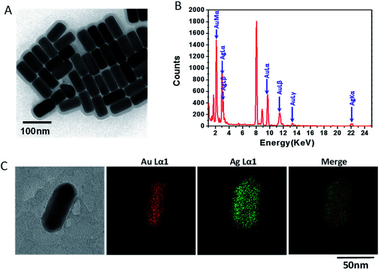
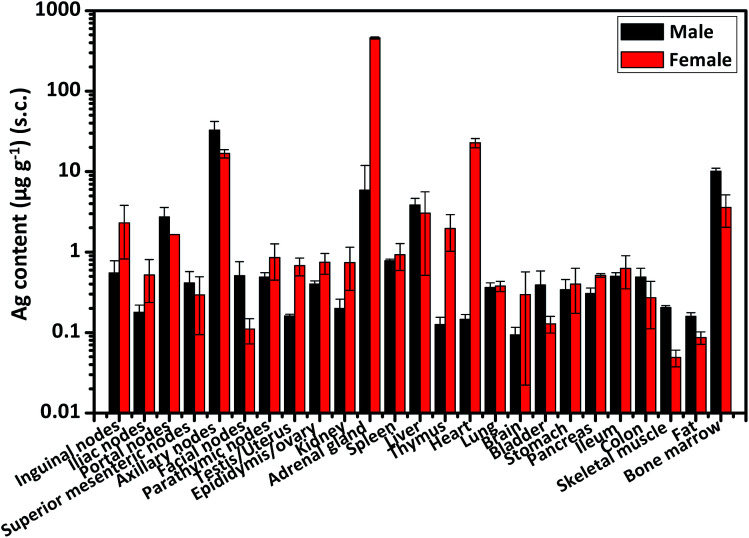
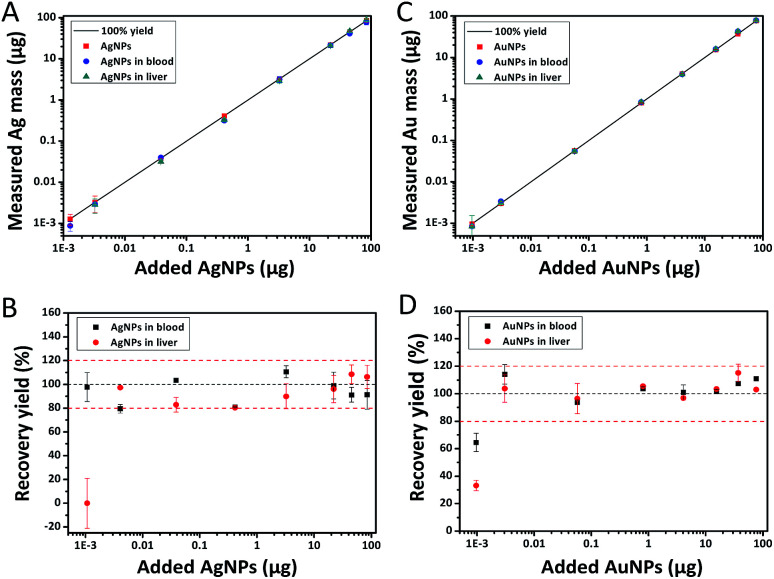
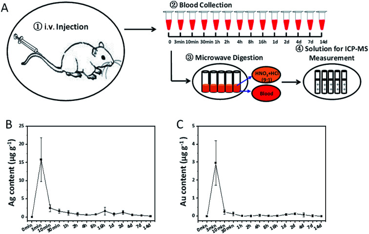
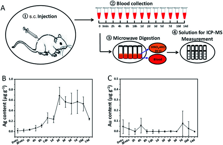
Along with the increasing applications of nanomaterials in medical fields, to know the systemic distribution of nanomaterials in the body through a precise method is required for the biosafety assessment of nanomaterials. In this study, we firstly have established a reliable inductively coupled plasma mass spectrometry (ICP-MS) method for concentration measurement of silver (Ag) and gold (Au) in biological tissues. Then, based on this method, the Ag or Au distribution in rat blood and almost all of the organs were analyzed after an i.v. or s.c. administration of Au@Ag NRs. Both the time-dependent contents of Ag and Au in blood and two pharmacokinetic models confirmed the rapid clearance of Ag from blood. At 24 h after i.v. injection, there was the highest level of Ag in liver, followed by portal nodes, spleen, lung, bone marrow and pancreas. In addition, we also found there were gender-related distributions of Ag and Au in some organs, especially after s.c. injection. Therefore, these more comprehensive and important results would give fundamental information for the biological risk assessment of nanomaterials.

摘要

随着纳米材料在医学领域的应用日益增加,通过精确方法了解纳米材料在体内的全身分布对于纳米材料的生物安全性评估至关重要。在本研究中,我们首先建立了一种可靠的电感耦合等离子体质谱(ICP-MS)方法,用于测量生物组织中银(Ag)和金(Au)的浓度。然后,基于该方法,在静脉注射或皮下注射Au@Ag纳米棒后,分析大鼠血液和几乎所有器官中的Ag或Au分布。血液中Ag和Au的时间依赖性含量以及两个药代动力学模型均证实Ag从血液中快速清除。静脉注射后24小时,肝脏中Ag的含量最高,其次是门静脉淋巴结、脾脏、肺、骨髓和胰腺。此外,我们还发现,在某些器官中,尤其是皮下注射后,Ag和Au存在与性别相关的分布。因此,这些更全面和重要的结果将为纳米材料的生物风险评估提供基础信息。

https://cdn.ncbi.nlm.nih.gov/pmc/blobs/9b93/9079266/9a3faa8ab28a/c8ra00044a-f6.jpg
https://cdn.ncbi.nlm.nih.gov/pmc/blobs/9b93/9079266/53706d8af689/c8ra00044a-f1.jpg
https://cdn.ncbi.nlm.nih.gov/pmc/blobs/9b93/9079266/f5ef0af71ff4/c8ra00044a-f2.jpg
https://cdn.ncbi.nlm.nih.gov/pmc/blobs/9b93/9079266/08c451f626c9/c8ra00044a-f3.jpg
https://cdn.ncbi.nlm.nih.gov/pmc/blobs/9b93/9079266/8cafd998c56f/c8ra00044a-f4.jpg
https://cdn.ncbi.nlm.nih.gov/pmc/blobs/9b93/9079266/3edac585564c/c8ra00044a-f5.jpg
https://cdn.ncbi.nlm.nih.gov/pmc/blobs/9b93/9079266/9a3faa8ab28a/c8ra00044a-f6.jpg

相似文献

1
Bio-distribution and bio-availability of silver and gold in rat tissues with silver/gold nanorod administration.银/金纳米棒给药后大鼠组织中银和金的生物分布及生物利用度
RSC Adv. 2018 Mar 29;8(22):12260-12268. doi: 10.1039/c8ra00044a. eCollection 2018 Mar 26.
2
Silver and gold nanoparticles characterization by SP-ICP-MS and AF4-FFF-MALS-UV-ICP-MS in human samples used for biomonitoring.通过单颗粒电感耦合等离子体质谱法(SP-ICP-MS)和不对称流场流分离-多角度激光散射-紫外-电感耦合等离子体质谱法(AF4-FFF-MALS-UV-ICP-MS)对用于生物监测的人体样本中的银和金纳米颗粒进行表征。
Talanta. 2020 Dec 1;220:121404. doi: 10.1016/j.talanta.2020.121404. Epub 2020 Jul 13.
3
Using gold nanorods core/silver shell nanostructures as model material to probe biodistribution and toxic effects of silver nanoparticles in mice.采用金纳米棒核/银壳纳米结构作为模型材料,来探究纳米银颗粒在小鼠体内的生物分布和毒效。
Nanotoxicology. 2014 Sep;8(6):686-96. doi: 10.3109/17435390.2013.822593. Epub 2013 Jul 29.
4
Interference of Steroidogenesis by Gold Nanorod Core/Silver Shell Nanostructures: Implications for Reproductive Toxicity of Silver Nanomaterials.金纳米棒核/银壳纳米结构对类固醇生成的干扰:对银纳米材料生殖毒性的影响。
Small. 2017 Mar;13(10). doi: 10.1002/smll.201602855. Epub 2016 Dec 23.
5
Tissue distribution of gold and silver after subacute intravenous injection of co-administered gold and silver nanoparticles of similar sizes.亚急性静脉注射相似大小的金和银纳米粒子缀合物后金和银在组织中的分布。
Arch Toxicol. 2018 Apr;92(4):1393-1405. doi: 10.1007/s00204-018-2173-4. Epub 2018 Feb 15.
6
Use of single particle inductively coupled plasma mass spectrometry for understanding the formation of bimetallic nanoparticles.利用单颗粒电感耦合等离子体质谱法了解双金属纳米颗粒的形成。
Talanta. 2022 Jan 1;236:122871. doi: 10.1016/j.talanta.2021.122871. Epub 2021 Sep 11.
7
[Determination of trace element silver in animal serum, tissues and organs by microwave digestion-ICP-MS].微波消解-电感耦合等离子体质谱法测定动物血清、组织和器官中的微量元素银
Guang Pu Xue Yu Guang Pu Fen Xi. 2014 Sep;34(9):2533-7.
8
The Bio-Persistence of Reversible Inflammatory, Histological Changes and Metabolic Profile Alterations in Rat Livers after Silver/Gold Nanorod Administration.银/金纳米棒给药后大鼠肝脏可逆性炎症、组织学变化及代谢谱改变的生物持久性
Nanomaterials (Basel). 2021 Oct 9;11(10):2656. doi: 10.3390/nano11102656.
9
[Determination of gold and silver in twenty plants from the copper mining area by inductively coupled plasma-mass spectrometry].[电感耦合等离子体质谱法测定铜矿区二十种植物中的金和银]
Guang Pu Xue Yu Guang Pu Fen Xi. 2012 May;32(5):1387-90.
10
Single-Dosed Genotoxicity Study of Gold Nanorod Core/Silver Shell Nanostructures by , Micronucleus, and Comet Assays.金纳米棒核/银壳纳米结构的单次给药遗传毒性研究:微核、彗星试验。
J Biomed Nanotechnol. 2018 Nov 1;14(11):1953-1964. doi: 10.1166/jbn.2018.2640.

引用本文的文献

1
Do We Know Enough About the Safety Profile of Silver Nanoparticles in Oncology? A Focus on Novel Methods and Approaches.我们对银纳米颗粒在肿瘤学中的安全性了解足够吗?聚焦于新方法和途径。
Int J Mol Sci. 2025 Jun 2;26(11):5344. doi: 10.3390/ijms26115344.
2
Transformation of Nano-Size Titanium Dioxide Particles in the Gastrointestinal Tract and Its Role in the Transfer of Nanoparticles through the Intestinal Barrier.纳米二氧化钛颗粒在胃肠道中的转化及其在纳米颗粒穿过肠屏障转移中的作用。
Int J Mol Sci. 2023 Oct 5;24(19):14911. doi: 10.3390/ijms241914911.
3
Bioluminescence Imaging and ICP-MS Associated with SPION as a Tool for Hematopoietic Stem and Progenitor Cells Homing and Engraftment Evaluation.

本文引用的文献

1
Silver nanoparticles: Significance of physicochemical properties and assay interference on the interpretation of in vitro cytotoxicity studies.银纳米颗粒:物理化学性质及检测干扰对体外细胞毒性研究解读的意义
Toxicol In Vitro. 2017 Feb;38:179-192. doi: 10.1016/j.tiv.2016.10.012. Epub 2016 Nov 3.
2
Silver Nanoparticle-Mediated Cellular Responses in Various Cell Lines: An in Vitro Model.银纳米颗粒介导的多种细胞系中的细胞反应:一种体外模型
Int J Mol Sci. 2016 Sep 22;17(10):1603. doi: 10.3390/ijms17101603.
3
Tissue distribution and acute toxicity of silver after single intravenous administration in mice: nano-specific and size-dependent effects.
生物发光成像和电感耦合等离子体质谱联用超顺磁性氧化铁纳米粒子作为评估造血干细胞归巢和植入的工具
Pharmaceutics. 2023 Mar 3;15(3):828. doi: 10.3390/pharmaceutics15030828.
4
Analytical Performance and Validation of a Reliable Method Based on Graphite Furnace Atomic Absorption Spectrometry for the Determination of Gold Nanoparticles in Biological Tissues.基于石墨炉原子吸收光谱法测定生物组织中纳米金颗粒的可靠方法的分析性能与验证
Nanomaterials (Basel). 2021 Dec 12;11(12):3370. doi: 10.3390/nano11123370.
5
Attomole-per Cell Atomic Mass Spectrometry Measurement of Platinum and Gold Drugs in Cultured Lung Cancer Cells.单细胞纳升级原子质谱法测定培养肺癌细胞中的铂类药物和金类药物。
Molecules. 2021 Dec 16;26(24):7627. doi: 10.3390/molecules26247627.
6
The Bio-Persistence of Reversible Inflammatory, Histological Changes and Metabolic Profile Alterations in Rat Livers after Silver/Gold Nanorod Administration.银/金纳米棒给药后大鼠肝脏可逆性炎症、组织学变化及代谢谱改变的生物持久性
Nanomaterials (Basel). 2021 Oct 9;11(10):2656. doi: 10.3390/nano11102656.
7
In Vivo Metabolic Response upon Exposure to Gold Nanorod Core/Silver Shell Nanostructures: Modulation of Inflammation and Upregulation of Dopamine.体内暴露于金纳米棒核/银壳纳米结构后的代谢反应:炎症调节和多巴胺上调。
Int J Mol Sci. 2020 Jan 8;21(2):384. doi: 10.3390/ijms21020384.
8
Effect of Gold Nanoparticle Size on Their Properties as Contrast Agents for Computed Tomography.金纳米颗粒尺寸对其作为计算机断层扫描造影剂性能的影响。
Sci Rep. 2019 Oct 17;9(1):14912. doi: 10.1038/s41598-019-50332-8.
9
Size-dependent cellular uptake and localization profiles of silver nanoparticles.银纳米粒子的尺寸依赖性细胞摄取和定位分布。
Int J Nanomedicine. 2019 Jun 7;14:4247-4259. doi: 10.2147/IJN.S201107. eCollection 2019.
小鼠单次静脉注射后银的组织分布及急性毒性:纳米特异性和尺寸依赖性效应
Part Fibre Toxicol. 2016 Feb 29;13:12. doi: 10.1186/s12989-016-0124-x.
4
Chemical basis of interactions between engineered nanoparticles and biological systems.工程纳米颗粒与生物系统之间相互作用的化学基础
Chem Rev. 2014 Aug 13;114(15):7740-81. doi: 10.1021/cr400295a. Epub 2014 Jun 13.
5
Radioactive 198Au-doped nanostructures with different shapes for in vivo analyses of their biodistribution, tumor uptake, and intratumoral distribution.具有不同形状的放射性198Au掺杂纳米结构用于体内生物分布、肿瘤摄取和瘤内分布分析。
ACS Nano. 2014 May 27;8(5):4385-94. doi: 10.1021/nn406258m. Epub 2014 Apr 30.
6
Systemic and immunotoxicity of silver nanoparticles in an intravenous 28 days repeated dose toxicity study in rats.静脉注射 28 天重复剂量毒性研究中银纳米粒子的全身和免疫毒性。
Biomaterials. 2013 Nov;34(33):8333-43. doi: 10.1016/j.biomaterials.2013.06.048. Epub 2013 Jul 22.
7
Using gold nanorods core/silver shell nanostructures as model material to probe biodistribution and toxic effects of silver nanoparticles in mice.采用金纳米棒核/银壳纳米结构作为模型材料,来探究纳米银颗粒在小鼠体内的生物分布和毒效。
Nanotoxicology. 2014 Sep;8(6):686-96. doi: 10.3109/17435390.2013.822593. Epub 2013 Jul 29.
8
Distribution, elimination, and toxicity of silver nanoparticles and silver ions in rats after 28-day oral exposure.口服暴露 28 天后大鼠体内银纳米颗粒和银离子的分布、消除和毒性。
ACS Nano. 2012 Aug 28;6(8):7427-42. doi: 10.1021/nn302649p. Epub 2012 Aug 9.
9
Acute toxic effects and gender-related biokinetics of silver nanoparticles following an intravenous injection in mice.静脉注射后小鼠体内银纳米颗粒的急性毒性效应及性别相关生物动力学
J Appl Toxicol. 2012 Nov;32(11):890-9. doi: 10.1002/jat.2742. Epub 2012 Apr 23.
10
Novel microfilaricidal activity of nanosilver.纳米银的新型微丝蚴杀灭活性。
Int J Nanomedicine. 2012;7:1023-30. doi: 10.2147/IJN.S28758. Epub 2012 Feb 22.