Black Kvar C L, Wang Yucai, Luehmann Hannah P, Cai Xin, Xing Wenxin, Pang Bo, Zhao Yongfeng, Cutler Cathy S, Wang Lihong V, Liu Yongjian, Xia Younan
Mallinckrodt Institute of Radiology, Washington University School of Medicine , St. Louis, Missouri 63110, United States.
ACS Nano. 2014 May 27;8(5):4385-94. doi: 10.1021/nn406258m. Epub 2014 Apr 30.
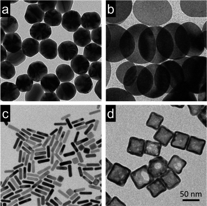
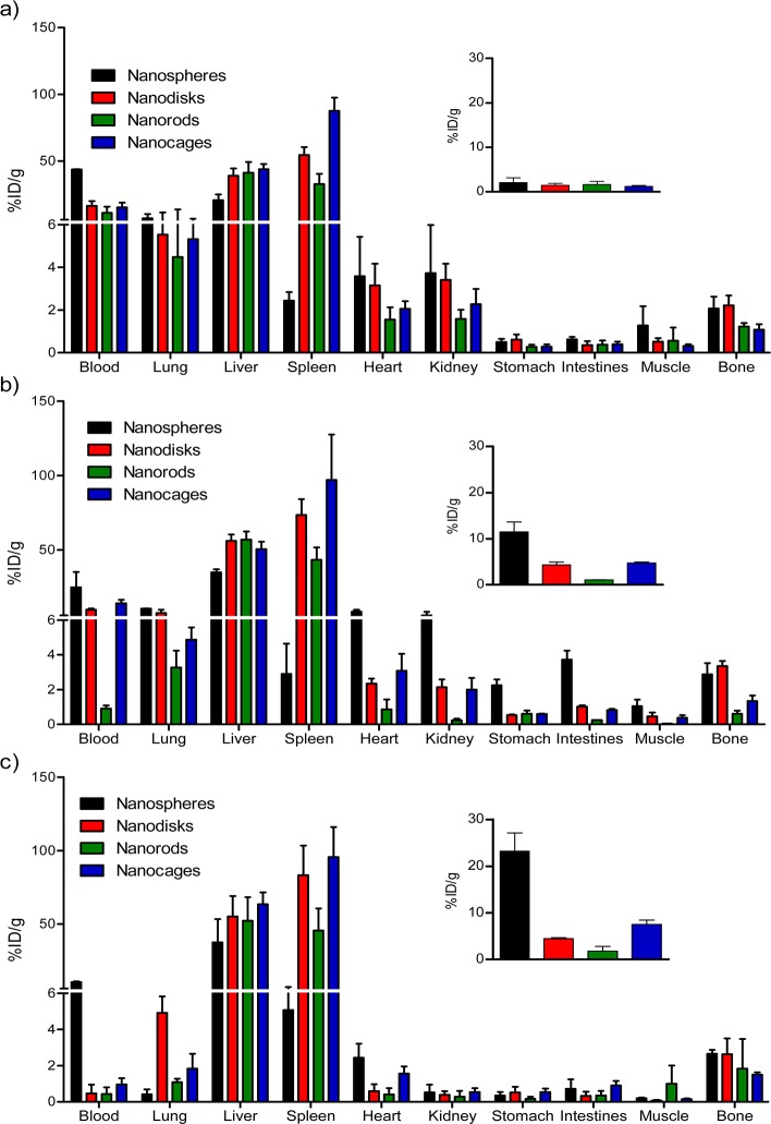
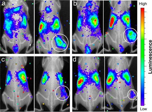
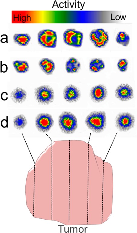
With Au nanocages as an example, we recently demonstrated that radioactive (198)Au could be incorporated into the crystal lattice of Au nanostructures for simple and reliable quantification of their in vivo biodistribution by measuring the γ radiation from (198)Au decay and for optical imaging by detecting the Cerenkov radiation. Here we extend the capability of this strategy to synthesize radioactive (198)Au nanostructures with a similar size but different shapes and then compare their biodistribution, tumor uptake, and intratumoral distribution using a murine EMT6 breast cancer model. Specifically, we investigated Au nanospheres, nanodisks, nanorods, and cubic nanocages. After PEGylation, an aqueous suspension of the radioactive Au nanostructures was injected into a tumor-bearing mouse intravenously, and their biodistribution was measured from the γ radiation while their tumor uptake was directly imaged using the Cerenkov radiation. Significantly higher tumor uptake was observed for the Au nanospheres and nanodisks relative to the Au nanorods and nanocages at 24 h postinjection. Furthermore, autoradiographic imaging was performed on thin slices of the tumor after excision to resolve the intratumoral distributions of the nanostructures. While both the Au nanospheres and nanodisks were only observed on the surfaces of the tumors, the Au nanorods and nanocages were distributed throughout the tumors.
以金纳米笼为例,我们最近证明,放射性(198)Au可以掺入金纳米结构的晶格中,通过测量(198)Au衰变产生的γ辐射来简单可靠地定量其体内生物分布,并通过检测切伦科夫辐射进行光学成像。在此,我们扩展了该策略的能力,合成了尺寸相似但形状不同的放射性(198)Au纳米结构,然后使用小鼠EMT6乳腺癌模型比较它们的生物分布、肿瘤摄取和肿瘤内分布。具体而言,我们研究了金纳米球、纳米盘、纳米棒和立方纳米笼。聚乙二醇化后,将放射性金纳米结构的水悬浮液静脉注射到荷瘤小鼠体内,通过γ辐射测量它们的生物分布,同时利用切伦科夫辐射直接成像它们的肿瘤摄取情况。注射后24小时,相对于金纳米棒和纳米笼,金纳米球和纳米盘的肿瘤摄取明显更高。此外,在切除肿瘤后对薄片进行放射自显影成像,以解析纳米结构在肿瘤内的分布。虽然金纳米球和纳米盘仅在肿瘤表面观察到,但金纳米棒和纳米笼分布在整个肿瘤中。