Labouta Hagar I, Sarsons Christopher, Kennard Jacob, Gomez-Garcia M Juliana, Villar Kenrick, Lee Hyungbok, Cramb David T, Rinker Kristina D
Department of Chemistry, Faculty of Science, University of Calgary Calgary Canada
Biomedical Engineering, University of Calgary Calgary Canada
RSC Adv. 2018 Jun 22;8(41):23027-23039. doi: 10.1039/c8ra03849j. eCollection 2018 Jun 21.
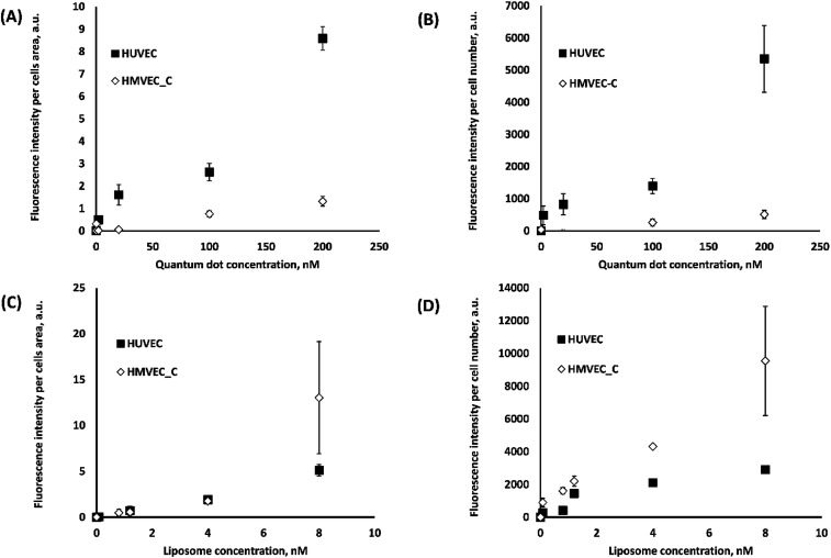
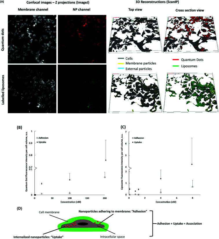
Despite years of excellent individual studies, the impact of nanoparticle (NP) cytotoxicity studies remains limited by inconsistent data collection and analysis. It is often unclear how exposure conditions can be used to determine cytotoxicity quantitatively. Discrepancies due to using different measurement conditions, readouts and controls to characterize NP interactions with cells lead to further challenges. To examine which parameters are critical in NP cytotoxicity studies, we have chosen to examine two NP types (liposomes and quantum dots) at different concentrations incubated with two primary vascular endothelial cells, HUVEC and HMVEC-C for a standard time of 24 h. We paid close attention to the effects of positive controls and cell association on interpretation of cytotoxicity data. Various cellular responses (ATP content, oxidative stress, mitochondrial toxicity, and phospholipidosis) were measured in parallel. Interestingly, cell association data varied significantly with the different image analyses. However, cytotoxicity responses could all be correlated with exposure concentration. Cell type did have an effect on cytotoxicity reports. Most significantly, NP cytotoxicity results varied with the inclusion or exclusion of positive controls. In the absence of positive controls, one tends to emphasize small changes in cell responses to NPs.
尽管多年来有出色的个体研究,但纳米颗粒(NP)细胞毒性研究的影响仍因数据收集和分析不一致而受到限制。通常不清楚如何利用暴露条件来定量确定细胞毒性。由于使用不同的测量条件、读数和对照来表征NP与细胞的相互作用而产生的差异导致了进一步的挑战。为了研究哪些参数在NP细胞毒性研究中至关重要,我们选择在标准的24小时内,用两种不同浓度的NP(脂质体和量子点)与两种原代血管内皮细胞HUVEC和HMVEC-C孵育。我们密切关注阳性对照和细胞结合对细胞毒性数据解释的影响。同时测量了各种细胞反应(ATP含量、氧化应激、线粒体毒性和磷脂蓄积)。有趣的是,不同的图像分析使细胞结合数据有显著差异。然而,细胞毒性反应都与暴露浓度相关。细胞类型确实对细胞毒性报告有影响。最重要的是,NP细胞毒性结果因是否包含阳性对照而有所不同。在没有阳性对照的情况下,人们往往会强调细胞对NP反应的微小变化。