Clemons T D, Bradshaw M, Toshniwal P, Chaudhari N, Stevenson A W, Lynch J, Fear M W, Wood F M, Iyer K Swaminathan
School of Molecular Sciences M313, The University of Western Australia 35 Stirling Hwy Crawley WA 6009 Australia
Fiona Wood Foundation and Burn Injury Research Unit, The University of Western Australia, M318 35 Stirling Hwy Crawley WA 6009 Australia.
RSC Adv. 2018 Mar 6;8(18):9661-9669. doi: 10.1039/c7ra12693j. eCollection 2018 Mar 5.
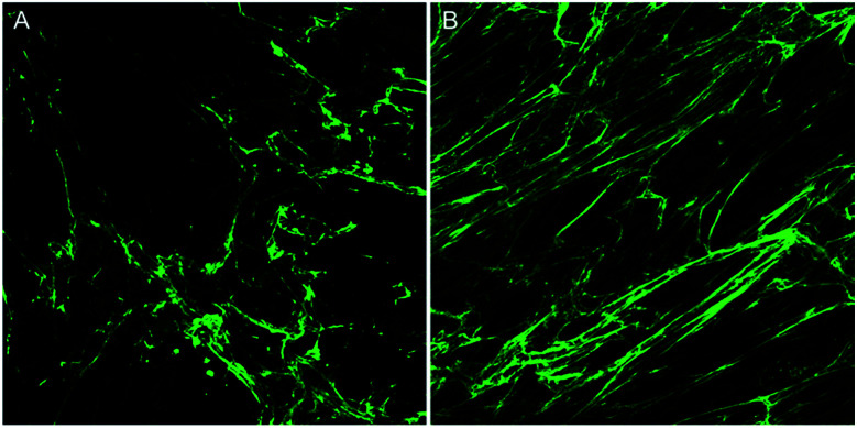
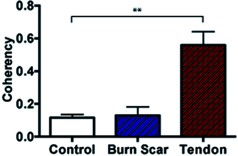
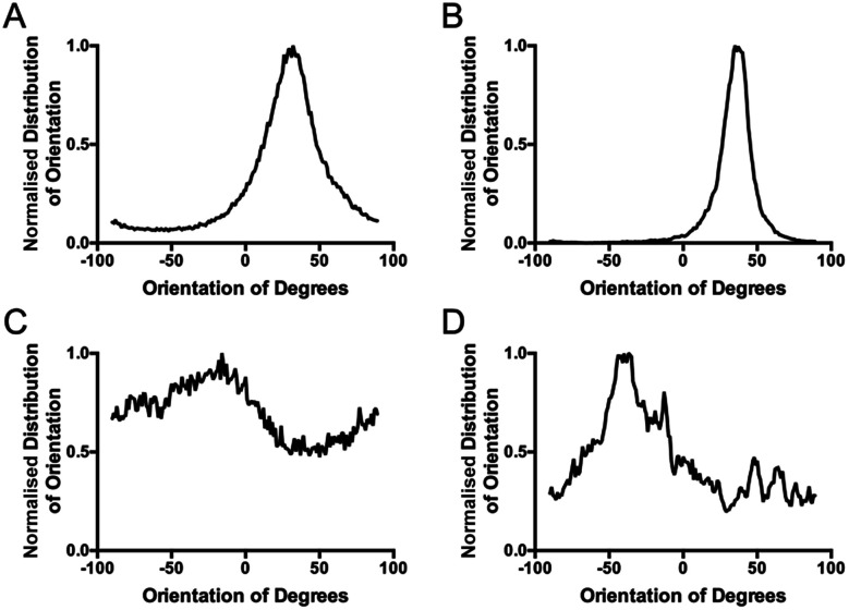
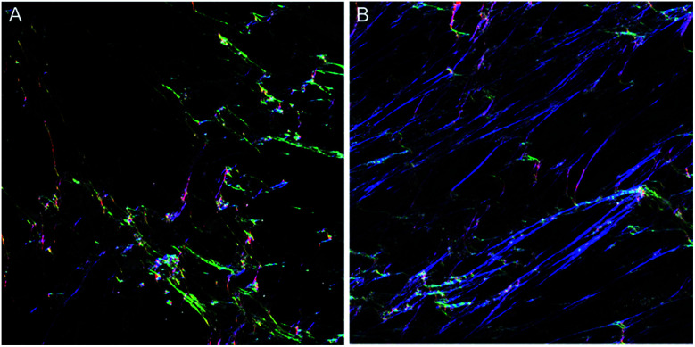
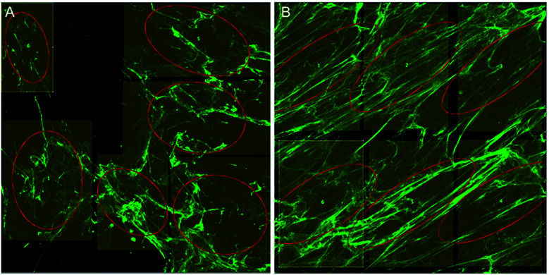
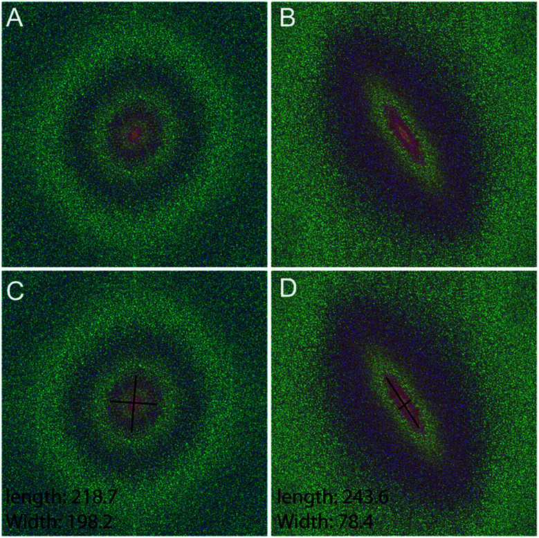
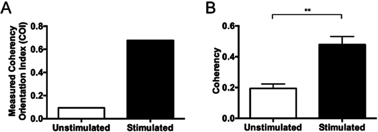
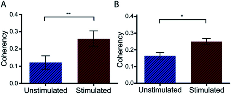
An important histological difference between normal, uninjured dermis and scar tissue such as that found in keloid scars is the pattern (morphological architecture) in which the collagen is deposited and arranged. In the uninjured dermis, collagen bundle architecture appears randomly organized (or in a basket weave formation), whereas in pathological conditions such as keloid scar tissue, collagen bundles are often found in whorls or in a hypotrophic scar collagen is more densely packed in a parallel configuration. In the case of skin, a scar disables the dermis, leaving it weaker, stiff and with a loss of optimal functionality. The absence of objective and quantifiable assessments of collagen orientation is a major bottleneck in monitoring progression of scar therapeutics. In this article, a novel quantitative approach for analyzing collagen orientation is reported. The methodology is demonstrated using collagen produced by cells in a model scar environment and examines collagen remodeling post-TGFβ stimulation . The method is shown to be reliable and effective in identifying significant coherency differences in the collagen deposited by human keloid scar cells. The technique is also compared for analysing collagen architecture in rat sections of normal, scarred skin and tendon tissue. Results demonstrate that the proposed computational method provides a fast and robust way of analyzing collagen orientation in a manner surpassing existing methods. This study establishes this methodology as a preliminary means of monitoring and in tissue treatment modalities which are expected to alter collagen morphology.
正常未受损真皮与瘢痕组织(如瘢痕疙瘩中的瘢痕组织)之间一个重要的组织学差异在于胶原蛋白沉积和排列的模式(形态结构)。在未受损的真皮中,胶原束结构呈现随机排列(或呈篮状编织结构),而在诸如瘢痕疙瘩瘢痕组织等病理状况下,胶原束常呈漩涡状排列,或者在增生性瘢痕中,胶原蛋白以平行排列的方式更为密集地堆积。就皮肤而言,瘢痕会使真皮功能受损,使其变得更脆弱、僵硬,且失去最佳功能。缺乏对胶原纤维排列方向的客观且可量化评估是监测瘢痕治疗进展的一个主要瓶颈。在本文中,报道了一种分析胶原纤维排列方向的新型定量方法。该方法通过在模型瘢痕环境中细胞产生的胶原蛋白进行演示,并研究转化生长因子β刺激后胶原重塑情况。结果表明,该方法在识别人类瘢痕疙瘩瘢痕细胞沉积的胶原蛋白中的显著相干性差异方面可靠且有效。该技术还用于分析正常、瘢痕化皮肤和肌腱组织的大鼠切片中的胶原结构。结果表明,所提出的计算方法提供了一种快速且强大的分析胶原纤维排列方向的方法,优于现有方法。本研究将该方法确立为监测以及在预期会改变胶原形态的组织治疗方式中的一种初步手段。