Kalaiarasi C, Manjula S, Kumaradhas P
Laboratory of Biocrystallography and Computational Molecular Biology, Department of Physics, Periyar University Salem-636 011 India
RSC Adv. 2019 Dec 10;9(69):40758-40771. doi: 10.1039/c9ra08607b. eCollection 2019 Dec 3.
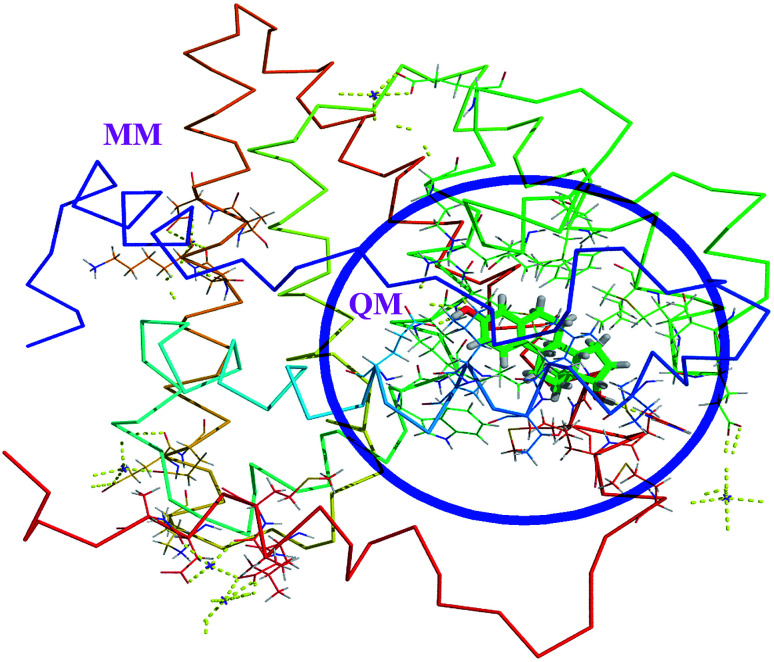
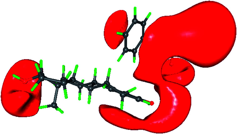
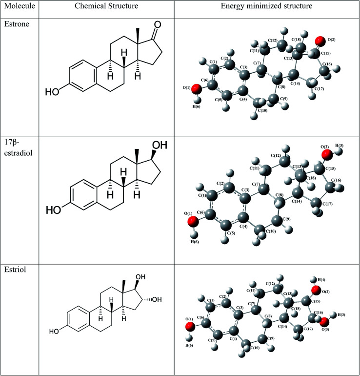
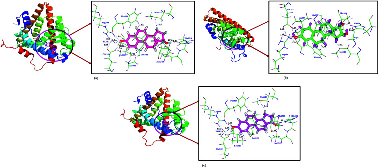
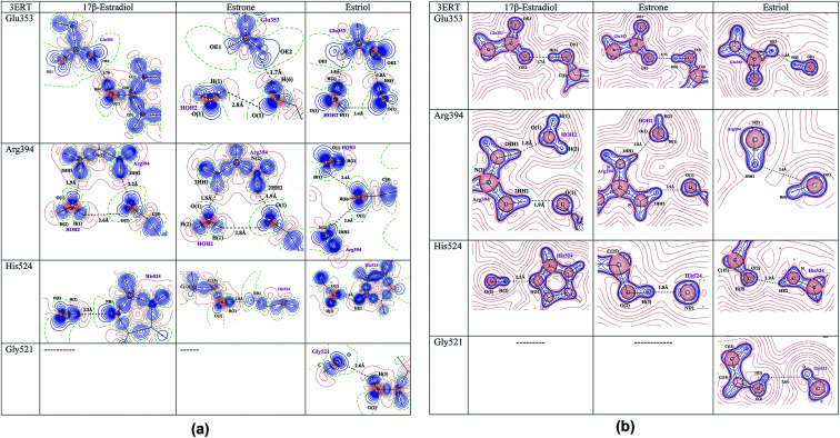
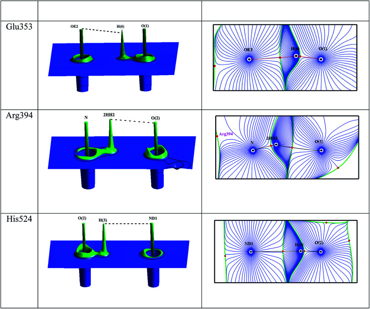
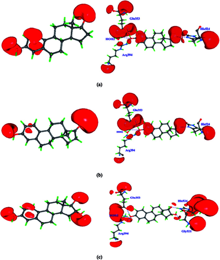
The ligand binding to protein and host-guest interactions are ubiquitous for molecular recognition. In drug design, the ligand binding to the active site of proteins is influenced by the charge density distribution and the electrostatic interactions of ligands and the nearby amino acids of the protein. The charge density analyses of ligand-protein complexes need accurate positions of hydrogen atoms and their valence electron distribution and the fine structure of proteins. Such information cannot be obtained from the conventional protein X-ray crystallography analysis in the resolution range of 1.5 to 3.5 Å. This can be realized from QM/MM based structure and charge density analysis of estrogens with the estrogen receptor. The charge density properties such as electron density, Laplacian of electron density and electrostatic properties of estrogens in the presence of active site amino acid residues have been determined and compared with the isolated estrogen molecules from theory and experimental. The present study reveals the chemical bonding nature of estrogen molecules and the strength of the intermolecular interactions in the active site of estrogen receptor, and also the importance of π⋯π interactions between the estrogens and Phe404 amino acid residue and protonation state of His524 amino acid residue have been identified using electrostatic potential maps. The difference in the electrostatic potential map of estrogens displays the hormone dependent actions of estrogen receptor. This method is very helpful to derive the charge density distribution of macromolecules to understand their biological recognition and interactions.
配体与蛋白质的结合以及主客体相互作用在分子识别中普遍存在。在药物设计中,配体与蛋白质活性位点的结合受配体以及蛋白质附近氨基酸的电荷密度分布和静电相互作用的影响。配体 - 蛋白质复合物的电荷密度分析需要氢原子的精确位置及其价电子分布以及蛋白质的精细结构。在1.5至3.5埃的分辨率范围内,从传统的蛋白质X射线晶体学分析中无法获得此类信息。这可以通过基于量子力学/分子力学(QM/MM)的雌激素与雌激素受体的结构和电荷密度分析来实现。已经确定了在活性位点氨基酸残基存在下雌激素的电荷密度性质,如电子密度、电子密度的拉普拉斯算子和静电性质,并与理论和实验得到的孤立雌激素分子进行了比较。本研究揭示了雌激素分子的化学键性质以及雌激素受体活性位点中分子间相互作用的强度,并且使用静电势图确定了雌激素与苯丙氨酸404氨基酸残基之间π⋯π相互作用以及组氨酸524氨基酸残基质子化状态的重要性。雌激素静电势图的差异显示了雌激素受体的激素依赖性作用。该方法对于推导大分子的电荷密度分布以理解其生物识别和相互作用非常有帮助。