Department of Radiology, Faculty of Medical Sciences, University of Fukui.
Department of Neurosurgery, Faculty of Medical Sciences, University of Fukui.
Magn Reson Med Sci. 2023 Jul 1;22(3):289-300. doi: 10.2463/mrms.mp.2020-0123. Epub 2022 May 10.
To verify whether arterial transit time (ATT) mapping can correct arterial spin labeling-cerebral blood flow (ASL-CBF) values and to verify whether ATT is a parameter that correlates with positron emission tomography (PET)-oxygen extraction fraction (OEF) and PET-mean transit time (MTT).
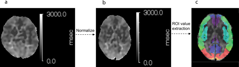
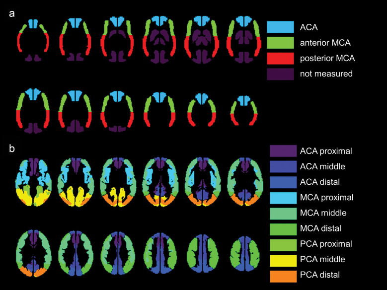
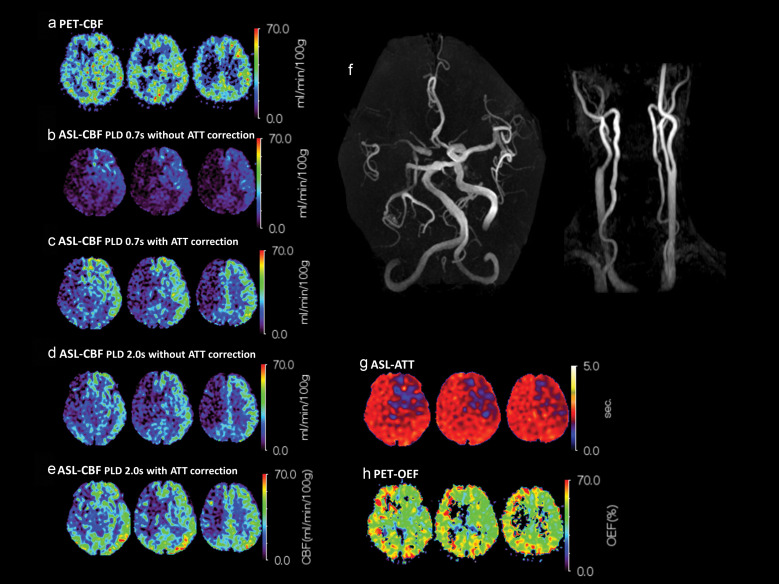
Eleven patients with unilateral major cerebral artery stenosis or occlusion underwent MRI and PET in the chronic or asymptomatic phase. ASL-MRI acquisitions were conducted with each of two post-label delay (PLD) settings (0.7s and 2.0s) using a pseudo-continuous ASL pulse sequence and 3D-spin echo spiral readout with vascular crusher gradient. ATT maps were obtained using a low-resolution pre-scan approach with five PLD settings. Using the ASL perfusion images and ATT mapping, ATT-corrected ASL-CBF images were obtained. Four kinds of ASL-CBF methods (PLD 0.7s with or without ATT correction and PLD 2.0s with or without ATT correction) were compared to PET-CBF, using vascular territory ROIs. ATT and OEF were compared for all ROIs, unaffected side ROIs, and affected side ROIs, respectively. ATT and MTT were compared by the ratio of the affected side to the unaffected side. Transit time-based ROIs were used for the comparison with ATT.
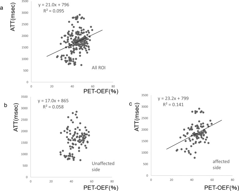
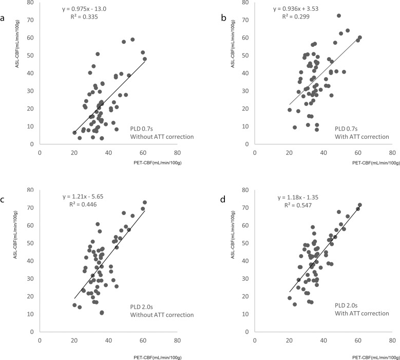
Comparing ASL-CBF and PET-CBF, the correlation was higher with ATT correction than without correction, and for a PLD of 2.0s compared with 0.7s. The best correlation was for PLD of 2.0s with ATT correction (R = 0.547). ROIs on the affected side showed a low but significant correlation between ATT and PET-OEF (R = 0.141). There was a low correlation between the ATT ratio and the MTT ratio (R = 0.133).
Low-resolution ATT correction may increase the accuracy of ASL-CBF measurements in patients with unilateral major cerebral artery stenosis or occlusion. In addition, ATT itself might have a potential role in detecting compromised hemodynamic state.
验证动脉渡越时间(ATT)图是否能校正动脉自旋标记-脑血流(ASL-CBF)值,并验证 ATT 是否是与正电子发射断层扫描(PET)-氧摄取分数(OEF)和 PET-平均通过时间(MTT)相关的参数。
11 例单侧大脑主要动脉狭窄或闭塞的患者在慢性或无症状期进行 MRI 和 PET 检查。使用伪连续 ASL 脉冲序列和带血管破碎机梯度的 3D 自旋回波螺旋读出,对两种后标记延迟(PLD)设置(0.7s 和 2.0s)进行 ASL-MRI 采集。使用具有五个 PLD 设置的低分辨率预扫描方法获得 ATT 图。使用 ASL 灌注图像和 ATT 映射,获得 ATT 校正的 ASL-CBF 图像。使用血管区域 ROI,比较了四种 ASL-CBF 方法(PLD 0.7s 加或不加 ATT 校正,以及 PLD 2.0s 加或不加 ATT 校正)与 PET-CBF。分别比较了所有 ROI、无影响侧 ROI 和受影响侧 ROI 的 ATT 和 OEF。分别比较了受累侧与未受累侧的 ATT 和 MTT 的比值。使用基于渡越时间的 ROI 与 ATT 进行比较。
与 PET-CBF 相比,校正 ATT 后 ASL-CBF 的相关性更高,而 2.0s 的 PLD 比 0.7s 的相关性更高。校正 ATT 后 2.0s 的 PLD 相关性最好(R=0.547)。受影响侧的 ROI 显示 ATT 与 PET-OEF 之间存在低但有统计学意义的相关性(R=0.141)。ATT 比值与 MTT 比值之间相关性较低(R=0.133)。
低分辨率 ATT 校正可能会提高单侧大脑主要动脉狭窄或闭塞患者 ASL-CBF 测量的准确性。此外,ATT 本身可能在检测血流动力学受损状态方面具有潜在作用。