• 文献检索
  • 文档翻译
  • 深度研究
  • 学术资讯
  • Suppr Zotero 插件Zotero 插件
  • 邀请有礼
  • 套餐&价格
  • 历史记录
应用&插件
Suppr Zotero 插件Zotero 插件浏览器插件Mac 客户端Windows 客户端微信小程序
定价
高级版会员购买积分包购买API积分包
服务
文献检索文档翻译深度研究API 文档MCP 服务
关于我们
关于 Suppr公司介绍联系我们用户协议隐私条款
关注我们

Suppr 超能文献

核心技术专利:CN118964589B侵权必究
粤ICP备2023148730 号-1Suppr @ 2026

文献检索

告别复杂PubMed语法,用中文像聊天一样搜索,搜遍4000万医学文献。AI智能推荐,让科研检索更轻松。

立即免费搜索

文件翻译

保留排版,准确专业,支持PDF/Word/PPT等文件格式,支持 12+语言互译。

免费翻译文档

深度研究

AI帮你快速写综述,25分钟生成高质量综述,智能提取关键信息,辅助科研写作。

立即免费体验

严重 COVID-19 患者发生肺纤维化时外周血单个核细胞及可溶性因子的变化。

Landscape of Peripheral Blood Mononuclear Cells and Soluble Factors in Severe COVID-19 Patients With Pulmonary Fibrosis Development.

机构信息

Department of Immunology, School of Basic Medical Sciences, Peking University. National Health Commission (NHC) Key Laboratory of Medical Immunology (Peking University), Beijing, China.

Beijing Youan Hospital, Capital Medical University, Beijing, China.

出版信息

Front Immunol. 2022 Apr 26;13:831194. doi: 10.3389/fimmu.2022.831194. eCollection 2022.

DOI:10.3389/fimmu.2022.831194
PMID:35558069
原文链接:https://pmc.ncbi.nlm.nih.gov/articles/PMC9088015/
Abstract

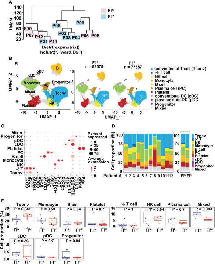
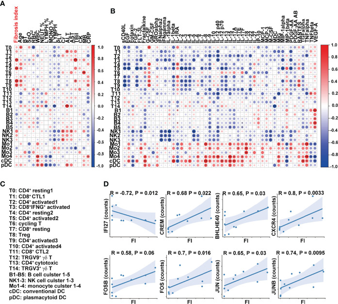
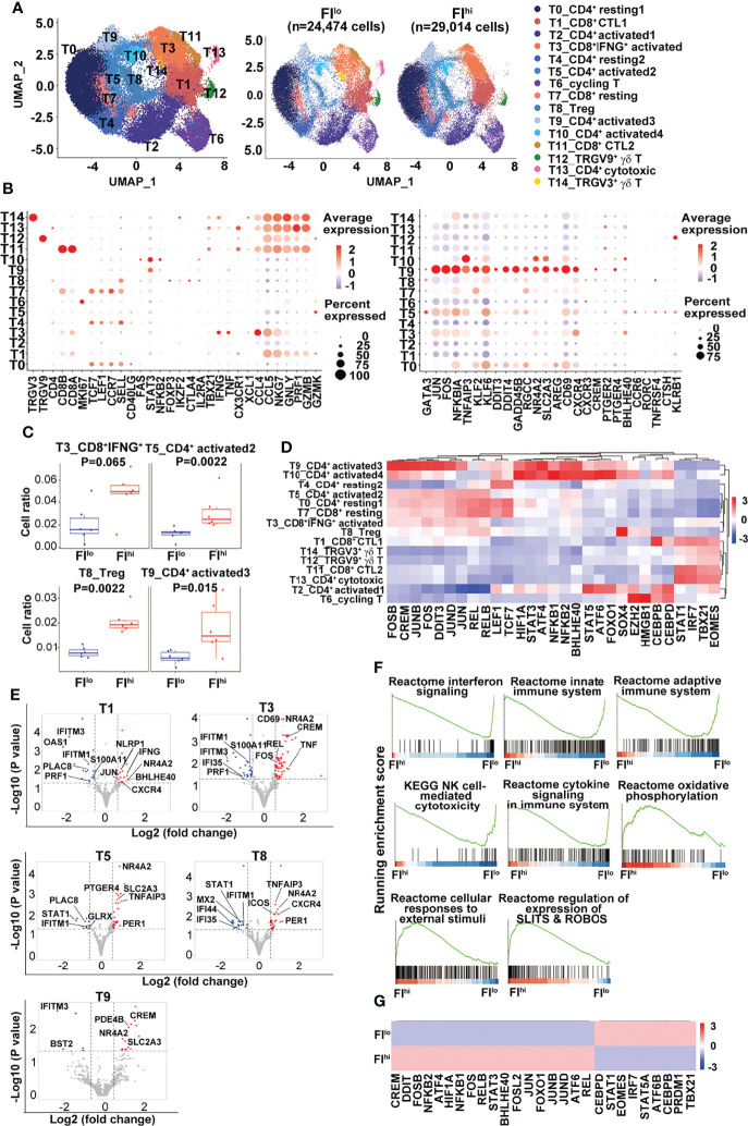
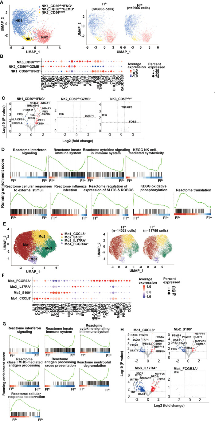
Resulting from severe inflammation and cell destruction, COVID-19 patients could develop pulmonary fibrosis (PF), which remains in the convalescent stage. Nevertheless, how immune response participates in the pathogenesis of PF progression is not well defined. To investigate that question, 12 patients with severe COVID-19 were included in the study. Peripheral mononuclear cell (PBMC) samples were collected shortly after their admission and proceeded for single-cell RNA sequencing (scRNA-seq). After 14 days of discharge, the patients were revisited for chest CT scan. PF index (FI) was computed by AI-assisted CT images. Patients were categorized into FI and FI based on median of FI. By scRNA-seq analysis, our data demonstrated that frequency of CD4+ activated T cells and Treg cells were approximately 3-fold higher in FI patients compared with FI ones ( < 0.034 for all). By dissecting the differentially expressed genes, we found an overall downregulation of IFN-responsive genes (, and ) and S100s alarmins (, etc.) in all T-cell clusters, and cytotoxicity-related genes (, and ) in CTLs and γδ T cells in the FI cohort, compared with FI subjects. The GSEA analysis illustrated decreased expression of genes enriched in IFN signaling, innate immune response, adaptive immune response in T cells, NK cells, and monocytes in FI patients compared with FI ones. In conclusion, these data indicated that the attenuated IFN-responsive genes and their related signaling pathways could be critical for PF progression in COVID-19 patients.

摘要

由于严重的炎症和细胞破坏,COVID-19 患者可能会发展为肺纤维化(PF),这种情况在康复期仍然存在。然而,免疫反应如何参与 PF 进展的发病机制尚不清楚。为了研究这个问题,本研究纳入了 12 名重症 COVID-19 患者。患者入院后不久采集外周血单核细胞(PBMC)样本,并进行单细胞 RNA 测序(scRNA-seq)。出院 14 天后,对患者进行胸部 CT 扫描复查。通过 AI 辅助 CT 图像计算 PF 指数(FI)。根据 FI 的中位数,患者被分为 FI 和 FI 两组。通过 scRNA-seq 分析,我们的数据表明,FI 患者的 CD4+活化 T 细胞和 Treg 细胞的频率比 FI 患者高约 3 倍(均<0.034)。通过对差异表达基因进行分析,我们发现 FI 组所有 T 细胞簇中 IFN 反应基因(、和)和 S100s 警报素(、等)总体下调,CTLs 和 γδ T 细胞中细胞毒性相关基因(、和)下调,与 FI 组相比。GSEA 分析表明,与 FI 组相比,FI 患者中 IFN 信号、固有免疫反应、T 细胞、NK 细胞和单核细胞适应性免疫反应相关基因的表达减少。综上所述,这些数据表明,IFN 反应基因及其相关信号通路的减弱可能是 COVID-19 患者 PF 进展的关键。

https://cdn.ncbi.nlm.nih.gov/pmc/blobs/20ce/9088015/94b5845717af/fimmu-13-831194-g005.jpg
https://cdn.ncbi.nlm.nih.gov/pmc/blobs/20ce/9088015/878fafa86a4c/fimmu-13-831194-g001.jpg
https://cdn.ncbi.nlm.nih.gov/pmc/blobs/20ce/9088015/6c8b4009bb6f/fimmu-13-831194-g002.jpg
https://cdn.ncbi.nlm.nih.gov/pmc/blobs/20ce/9088015/e30465524660/fimmu-13-831194-g003.jpg
https://cdn.ncbi.nlm.nih.gov/pmc/blobs/20ce/9088015/62eaa341bdbf/fimmu-13-831194-g004.jpg
https://cdn.ncbi.nlm.nih.gov/pmc/blobs/20ce/9088015/94b5845717af/fimmu-13-831194-g005.jpg

相似文献

1
Landscape of Peripheral Blood Mononuclear Cells and Soluble Factors in Severe COVID-19 Patients With Pulmonary Fibrosis Development.严重 COVID-19 患者发生肺纤维化时外周血单个核细胞及可溶性因子的变化。
Front Immunol. 2022 Apr 26;13:831194. doi: 10.3389/fimmu.2022.831194. eCollection 2022.
2
Single-cell RNA sequencing of peripheral blood reveals immune cell dysfunction in premature ovarian insufficiency.外周血单细胞 RNA 测序揭示卵巢早衰患者免疫细胞功能障碍。
Front Endocrinol (Lausanne). 2023 May 8;14:1129657. doi: 10.3389/fendo.2023.1129657. eCollection 2023.
3
High-dimensional comparison of monocytes and T cells in post-COVID and idiopathic pulmonary fibrosis.新冠后和特发性肺纤维化中单核细胞和 T 细胞的高维比较。
Front Immunol. 2024 Jan 16;14:1308594. doi: 10.3389/fimmu.2023.1308594. eCollection 2023.
4
Integrating single-cell sequencing data with GWAS summary statistics reveals CD16+monocytes and memory CD8+T cells involved in severe COVID-19.单细胞测序数据与 GWAS 汇总统计数据的整合揭示了 CD16+单核细胞和记忆 CD8+T 细胞参与严重 COVID-19。
Genome Med. 2022 Feb 17;14(1):16. doi: 10.1186/s13073-022-01021-1.
5
The Transient IFN Response and the Delay of Adaptive Immunity Feature the Severity of COVID-19.瞬时 IFN 反应和适应性免疫的延迟是 COVID-19 严重程度的特征。
Front Immunol. 2022 Jan 14;12:816745. doi: 10.3389/fimmu.2021.816745. eCollection 2021.
6
Dynamic blood single-cell immune responses in patients with COVID-19.COVID-19 患者的动态血液单细胞免疫反应。
Signal Transduct Target Ther. 2021 Mar 6;6(1):110. doi: 10.1038/s41392-021-00526-2.
7
Immunological Profiling of COVID-19 Patients with Pulmonary Sequelae.COVID-19 患者肺部后遗症的免疫特征分析。
mBio. 2021 Oct 26;12(5):e0159921. doi: 10.1128/mBio.01599-21. Epub 2021 Sep 7.
8
Identification of Distinct Immune Cell Subsets Associated With Asymptomatic Infection, Disease Severity, and Viral Persistence in COVID-19 Patients.鉴定与 COVID-19 患者无症状感染、疾病严重程度和病毒持续存在相关的不同免疫细胞亚群。
Front Immunol. 2022 Feb 22;13:812514. doi: 10.3389/fimmu.2022.812514. eCollection 2022.
9
Comparative transcriptome analysis of the transcriptional heterogeneity in different IgM cell subsets from peripheral blood of Nile tilapia (Oreochromis niloticus).比较尼罗罗非鱼(Oreochromis niloticus)外周血中不同 IgM 细胞亚群转录异质性的转录组分析。
Fish Shellfish Immunol. 2019 Oct;93:612-622. doi: 10.1016/j.fsi.2019.08.023. Epub 2019 Aug 10.
10
Lower Circulating Interferon-Gamma Is a Risk Factor for Lung Fibrosis in COVID-19 Patients.循环中较低水平的干扰素-γ是 COVID-19 患者肺纤维化的一个风险因素。
Front Immunol. 2020 Sep 29;11:585647. doi: 10.3389/fimmu.2020.585647. eCollection 2020.

引用本文的文献

1
Extracellular matrix remodelling pathway in peripheral blood mononuclear cells from severe COVID-19 patients: an explorative study.严重 COVID-19 患者外周血单个核细胞细胞外基质重塑途径:一项探索性研究。
Front Immunol. 2024 Jun 18;15:1379570. doi: 10.3389/fimmu.2024.1379570. eCollection 2024.
2
The deciphering of the immune cells and marker signature in COVID-19 pathogenesis: An update.解析 COVID-19 发病机制中的免疫细胞和标志物特征:最新进展。
J Med Virol. 2022 Nov;94(11):5128-5148. doi: 10.1002/jmv.28000. Epub 2022 Jul 23.
3
Prior Vaccination Exceeds Prior Infection in Eliciting Innate and Humoral Immune Responses in Omicron Infected Outpatients.

本文引用的文献

1
COVID-19 Lung Pathogenesis in SARS-CoV-2 Autopsy Cases.COVID-19 肺脏病理学:基于 SARS-CoV-2 尸检病例的研究。
Front Immunol. 2021 Oct 4;12:735922. doi: 10.3389/fimmu.2021.735922. eCollection 2021.
2
Endoplasmic Reticulum Quality Control in Immune Cells.免疫细胞中的内质网质量控制
Front Cell Dev Biol. 2021 Sep 29;9:740653. doi: 10.3389/fcell.2021.740653. eCollection 2021.
3
Early IFN-α signatures and persistent dysfunction are distinguishing features of NK cells in severe COVID-19.早期 IFN-α 特征和持续功能障碍是严重 COVID-19 中 NK 细胞的显著特征。
与既往感染相比,奥密克戎感染的门诊患者接种疫苗可引发更强的固有免疫和体液免疫应答。
Front Immunol. 2022 Jun 15;13:916686. doi: 10.3389/fimmu.2022.916686. eCollection 2022.
Immunity. 2021 Nov 9;54(11):2650-2669.e14. doi: 10.1016/j.immuni.2021.09.002. Epub 2021 Sep 4.
4
Confronting false discoveries in single-cell differential expression.单细胞差异表达中虚假发现的应对策略。
Nat Commun. 2021 Sep 28;12(1):5692. doi: 10.1038/s41467-021-25960-2.
5
From ARDS to pulmonary fibrosis: the next phase of the COVID-19 pandemic?从急性呼吸窘迫综合征到肺纤维化:COVID-19 大流行的下一阶段?
Transl Res. 2022 Mar;241:13-24. doi: 10.1016/j.trsl.2021.09.001. Epub 2021 Sep 20.
6
Lung Fibrosis after COVID-19: Treatment Prospects.新冠病毒感染后肺纤维化:治疗前景
Pharmaceuticals (Basel). 2021 Aug 17;14(8):807. doi: 10.3390/ph14080807.
7
The cAMP responsive element modulator (CREM) is a regulator of CD4 T cell function.环磷腺苷反应元件结合蛋白(CREM)是调节 CD4 T 细胞功能的一种调节蛋白。
Biol Chem. 2021 Aug 26;402(12):1591-1596. doi: 10.1515/hsz-2021-0249. Print 2021 Nov 25.
8
Tissue factor upregulation is associated with SARS-CoV-2 in the lungs of COVID-19 patients.组织因子上调与 COVID-19 患者肺部的 SARS-CoV-2 有关。
J Thromb Haemost. 2021 Sep;19(9):2268-2274. doi: 10.1111/jth.15451. Epub 2021 Jul 18.
9
The derepression of transposable elements in lung cells is associated with the inflammatory response and gene activation in idiopathic pulmonary fibrosis.肺细胞中转座元件的去抑制与特发性肺纤维化中的炎症反应和基因激活有关。
Mob DNA. 2021 Jun 9;12(1):14. doi: 10.1186/s13100-021-00241-3.
10
3-month, 6-month, 9-month, and 12-month respiratory outcomes in patients following COVID-19-related hospitalisation: a prospective study.COVID-19 相关住院患者的 3 个月、6 个月、9 个月和 12 个月呼吸结局:一项前瞻性研究。
Lancet Respir Med. 2021 Jul;9(7):747-754. doi: 10.1016/S2213-2600(21)00174-0. Epub 2021 May 5.