Brandenburg Leonard Simon, Schlager Stefan, Harzig Lara Sophie, Steybe David, Rothweiler René Marcel, Burkhardt Felix, Spies Benedikt Christopher, Georgii Joachim, Metzger Marc Christian
Medical Center-University of Freiburg, Center for Dental Medicine, Department of Oral and Maxillofacial Surgery, Faculty of Medicine, University of Freiburg, Hugstetter Str. 55, 79106 Freiburg, Germany.
Medical Center-University of Freiburg, Center for Dental Medicine, Department of Prosthetic Dentistry, Faculty of Medicine, University of Freiburg, Hugstetter Str. 55, 79106 Freiburg, Germany.
J Clin Med. 2022 Apr 24;11(9):2383. doi: 10.3390/jcm11092383.
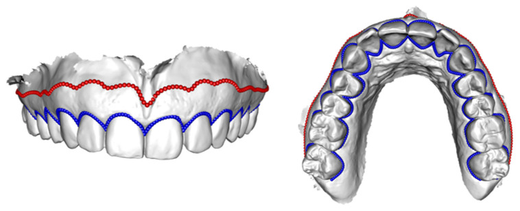
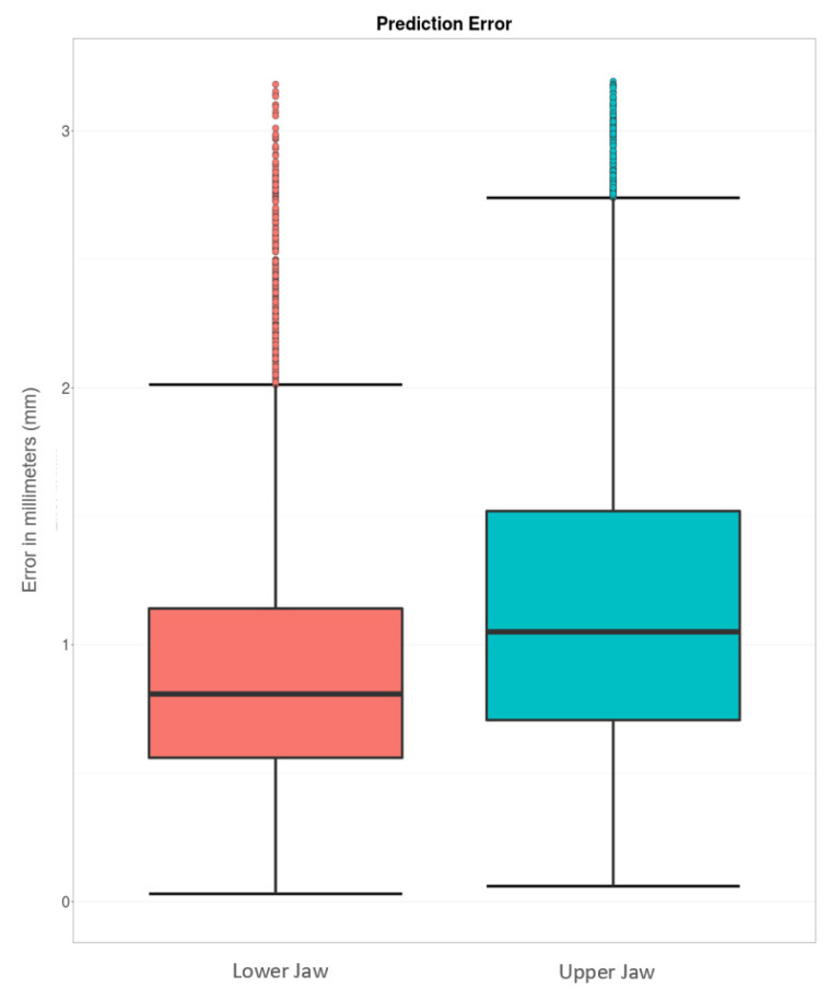
Adequate soft-tissue dimensions have been shown to be crucial for the long-term success of dental implants. To date, there is evidence that placement of dental implants should only be conducted in an area covered with attached gingiva. Modern implant planning software does not visualize soft-tissue dimensions. This study aims to calculate the course of the mucogingival borderline (MG-BL) using statistical shape models (SSM). Visualization of the MG-BL allows the practitioner to consider the soft tissue supply during implant planning. To deploy an SSM of the MG-BL, healthy individuals were examined and the intra-oral anatomy was captured using an intra-oral scanner (IOS). The empirical anatomical data was superimposed and analyzed by principal component analysis. Using a Leave-One-Out Cross Validation (LOOCV), the prediction of the SSM was compared with the original anatomy extracted from IOS. The median error for MG-BL reconstruction was 1.06 mm (0.49-2.15 mm) and 0.81 mm (0.38-1.54 mm) for the maxilla and mandible, respectively. While this method forgoes any technical work or additional patient examination, it represents an effective and digital method for the depiction of soft-tissue dimensions. To achieve clinical applicability, a higher number of datasets has to be implemented in the SSM.
充足的软组织尺寸已被证明对牙种植体的长期成功至关重要。迄今为止,有证据表明牙种植体的植入应仅在附着龈覆盖的区域进行。现代种植规划软件无法可视化软组织尺寸。本研究旨在使用统计形状模型(SSM)计算黏膜牙龈边界(MG-BL)的走向。MG-BL的可视化使从业者在种植规划过程中能够考虑软组织供应情况。为了构建MG-BL的SSM,对健康个体进行了检查,并使用口腔内扫描仪(IOS)获取口腔内解剖结构。将经验性解剖数据进行叠加,并通过主成分分析进行分析。使用留一法交叉验证(LOOCV),将SSM的预测结果与从IOS提取的原始解剖结构进行比较。上颌和下颌MG-BL重建的中位误差分别为1.06毫米(0.49 - 2.15毫米)和0.81毫米(0.38 - 1.54毫米)。虽然这种方法无需任何技术工作或额外的患者检查,但它是一种描绘软组织尺寸的有效数字方法。为了实现临床适用性,必须在SSM中纳入更多的数据集。