• 文献检索
  • 文档翻译
  • 深度研究
  • 学术资讯
  • Suppr Zotero 插件Zotero 插件
  • 邀请有礼
  • 套餐&价格
  • 历史记录
应用&插件
Suppr Zotero 插件Zotero 插件浏览器插件Mac 客户端Windows 客户端微信小程序
定价
高级版会员购买积分包购买API积分包
服务
文献检索文档翻译深度研究API 文档MCP 服务
关于我们
关于 Suppr公司介绍联系我们用户协议隐私条款
关注我们

Suppr 超能文献

核心技术专利:CN118964589B侵权必究
粤ICP备2023148730 号-1Suppr @ 2026

文献检索

告别复杂PubMed语法,用中文像聊天一样搜索,搜遍4000万医学文献。AI智能推荐,让科研检索更轻松。

立即免费搜索

文件翻译

保留排版,准确专业,支持PDF/Word/PPT等文件格式,支持 12+语言互译。

免费翻译文档

深度研究

AI帮你快速写综述,25分钟生成高质量综述,智能提取关键信息,辅助科研写作。

立即免费体验

眼表疾病临床体征与不适不一致的研究参与者的角膜共聚焦显微镜特征及泪液分子谱

Corneal Confocal Microscopy Features and Tear Molecular Profile in Study Participants with Discordance between Ocular Surface Disease Clinical Signs and Discomfort.

作者信息

D'Souza Sharon, Shetty Rohit, Nair Archana Padmanabhan, Agrawal Ruchika, Dickman Mor M, Khamar Pooja, Nuijts Rudy M M A, Ghosh Arkasubhra, Sethu Swaminathan

机构信息

Department of Cornea and Refractive Surgery, Narayana Nethralaya, Bangalore 560010, India.

GROW Research Laboratory, Narayana Nethralaya Foundation, Bangalore 560099, India.

出版信息

J Clin Med. 2022 Apr 25;11(9):2407. doi: 10.3390/jcm11092407.

DOI:10.3390/jcm11092407
PMID:35566533
原文链接:https://pmc.ncbi.nlm.nih.gov/articles/PMC9099769/
Abstract

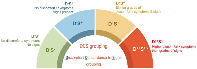
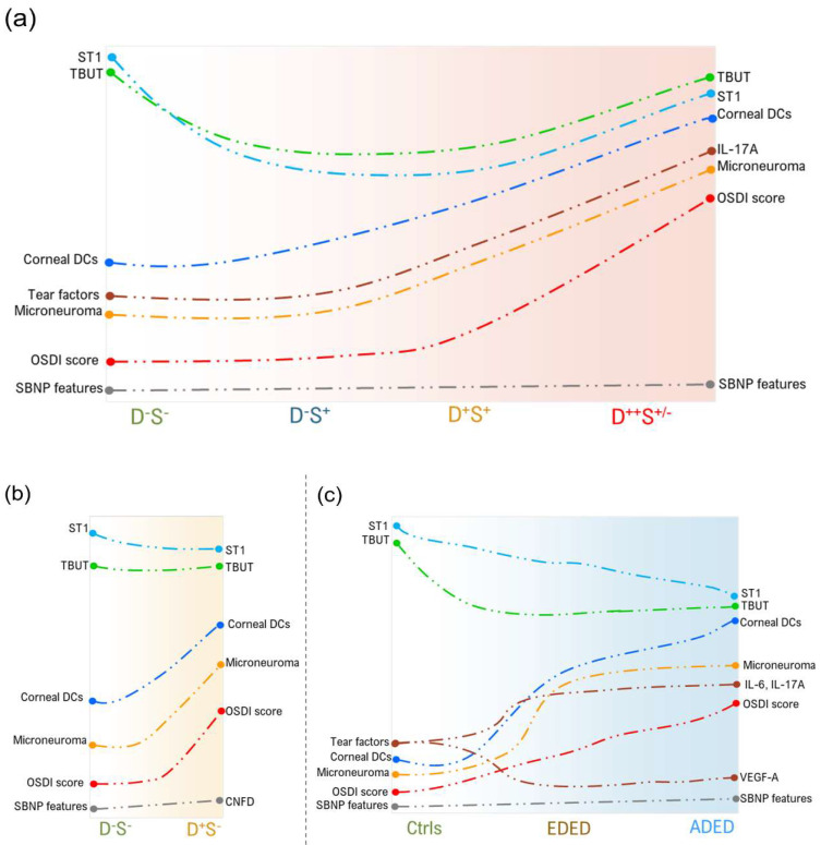
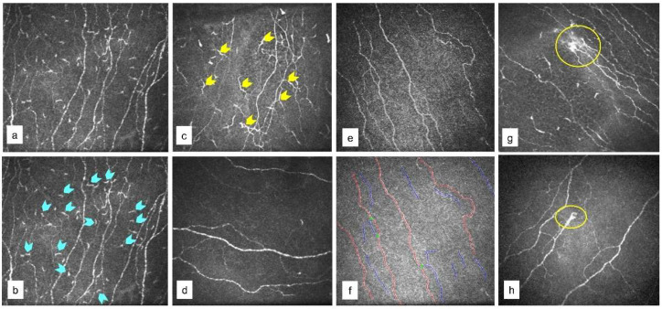
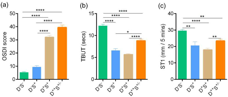
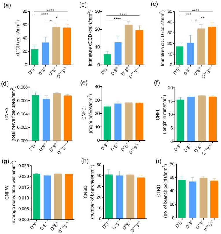
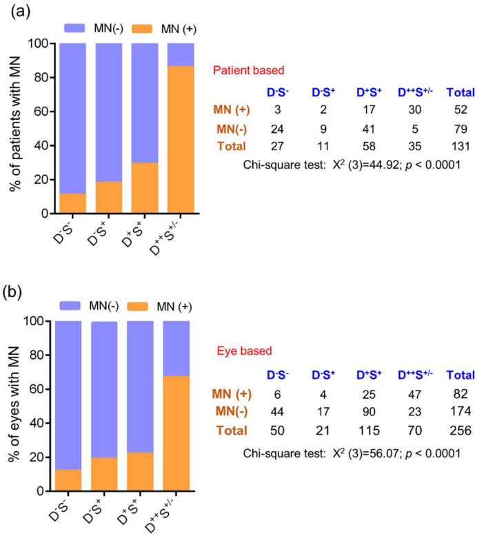
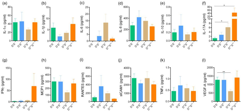
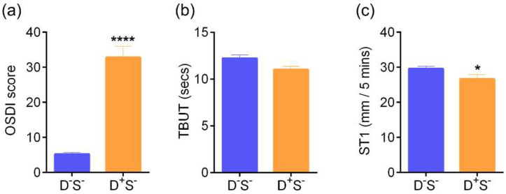
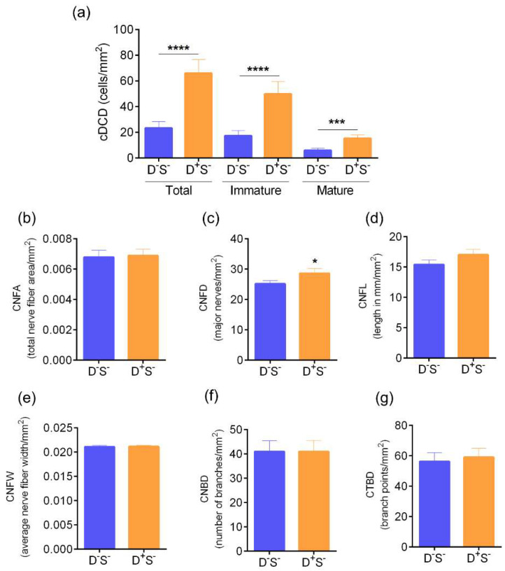
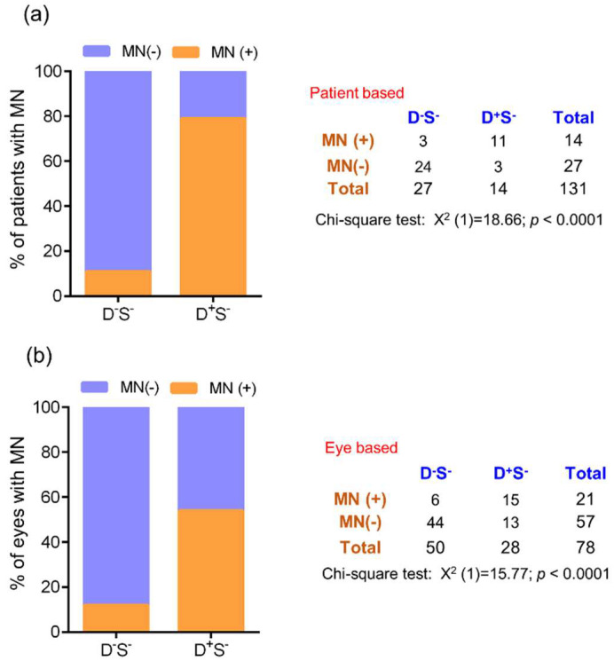
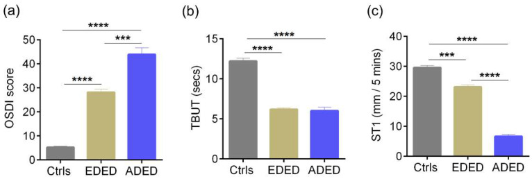
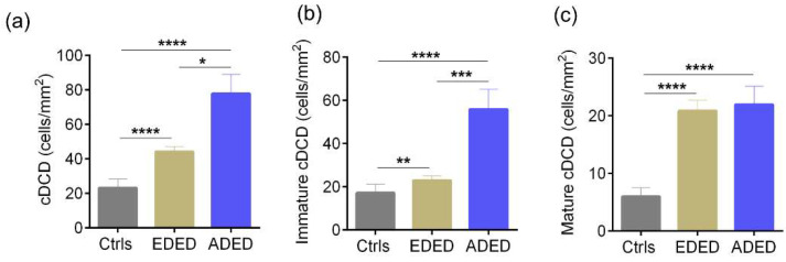
Various ocular surface conditions such as dry eye disease can present with severe discomfort and pain. However, it is clinically challenging to establish etiology and prescribe correct treatment in patients who have a lot of discordance between symptoms and signs. To understand the basis of such discordance, we stratified subjects with ocular surface pain based on concordance between the severity of signs and symptoms and evaluated corneal structural features and tear molecular factors. All subjects underwent slit lamp examination, dry eye evaluation, and ocular surface disease index (OSDI) scoring. Subjects were stratified into group 1—without symptoms or clinical signs; group 2—without symptoms but with signs; group 3—with similar severity of symptoms and signs; and group 4—with symptom severity greater than that of the signs. Laser scanning in vivo confocal microscopy (IVCM) and tear fluid analysis for soluble factors by multiplex ELISA was performed for all subjects. Patients with a higher grade of symptoms and signs showed increased corneal dendritic cell (cDC) density (p < 0.05) which was more pronounced in subjects with discordance between the symptoms and signs (group 4). A significantly higher proportion of microneuroma-like structures and cDC were observed in group 4. IL-17A levels were significantly elevated in the tears of subjects with more discomfort. Our results demonstrate that corneal IVCM and the measurement of tear film factors can help clinicians improve diagnosis and treatment choice. Stratifying patients with ocular surface discomfort on the basis of discordance between symptoms and clinical signs may help identify patients who need additional adjunctive targeted therapy to resolve their condition.

摘要

各种眼表疾病,如干眼症,可导致严重不适和疼痛。然而,对于症状与体征之间存在诸多不一致的患者,临床上要确定病因并开出正确的治疗方案具有挑战性。为了解这种不一致的原因,我们根据体征和症状的严重程度一致性,对眼表疼痛的受试者进行分层,并评估角膜结构特征和泪液分子因素。所有受试者均接受裂隙灯检查、干眼评估和眼表疾病指数(OSDI)评分。受试者被分为1组——无症状或临床体征;2组——无症状但有体征;3组——症状和体征严重程度相似;4组——症状严重程度大于体征。对所有受试者进行了激光扫描活体共聚焦显微镜检查(IVCM)和通过多重ELISA对泪液中的可溶性因子进行分析。症状和体征分级较高的患者角膜树突状细胞(cDC)密度增加(p < 0.05),在症状与体征不一致的受试者(4组)中更为明显。在4组中观察到微神经瘤样结构和cDC的比例显著更高。不适程度较高的受试者泪液中IL-17A水平显著升高。我们的结果表明,角膜IVCM和泪膜因子的测量可以帮助临床医生改善诊断和治疗选择。根据症状与临床体征之间的不一致对眼表不适患者进行分层,可能有助于识别需要额外辅助靶向治疗来解决其病情的患者。

https://cdn.ncbi.nlm.nih.gov/pmc/blobs/342f/9099769/00068292dad4/jcm-11-02407-g012.jpg
https://cdn.ncbi.nlm.nih.gov/pmc/blobs/342f/9099769/8a438b70c497/jcm-11-02407-g001.jpg
https://cdn.ncbi.nlm.nih.gov/pmc/blobs/342f/9099769/c5d2700e2e2a/jcm-11-02407-g002.jpg
https://cdn.ncbi.nlm.nih.gov/pmc/blobs/342f/9099769/1ca8ad35e0b5/jcm-11-02407-g003.jpg
https://cdn.ncbi.nlm.nih.gov/pmc/blobs/342f/9099769/c28d6c951cdc/jcm-11-02407-g004.jpg
https://cdn.ncbi.nlm.nih.gov/pmc/blobs/342f/9099769/e87c0416d8a1/jcm-11-02407-g005.jpg
https://cdn.ncbi.nlm.nih.gov/pmc/blobs/342f/9099769/77e4df22539b/jcm-11-02407-g006.jpg
https://cdn.ncbi.nlm.nih.gov/pmc/blobs/342f/9099769/f3491b40c6e1/jcm-11-02407-g007.jpg
https://cdn.ncbi.nlm.nih.gov/pmc/blobs/342f/9099769/1c46c80c829e/jcm-11-02407-g008.jpg
https://cdn.ncbi.nlm.nih.gov/pmc/blobs/342f/9099769/ce3b368c1c84/jcm-11-02407-g009.jpg
https://cdn.ncbi.nlm.nih.gov/pmc/blobs/342f/9099769/cb4600563d65/jcm-11-02407-g010.jpg
https://cdn.ncbi.nlm.nih.gov/pmc/blobs/342f/9099769/f1789c4b3798/jcm-11-02407-g011.jpg
https://cdn.ncbi.nlm.nih.gov/pmc/blobs/342f/9099769/00068292dad4/jcm-11-02407-g012.jpg

相似文献

1
Corneal Confocal Microscopy Features and Tear Molecular Profile in Study Participants with Discordance between Ocular Surface Disease Clinical Signs and Discomfort.眼表疾病临床体征与不适不一致的研究参与者的角膜共聚焦显微镜特征及泪液分子谱
J Clin Med. 2022 Apr 25;11(9):2407. doi: 10.3390/jcm11092407.
2
Dysregulated Tear Fluid Nociception-Associated Factors, Corneal Dendritic Cell Density, and Vitamin D Levels in Evaporative Dry Eye.异常的泪液伤害感受相关因子、角膜树突状细胞密度和蒸发性干眼的维生素 D 水平。
Invest Ophthalmol Vis Sci. 2019 Jun 3;60(7):2532-2542. doi: 10.1167/iovs.19-26914.
3
Corneal neuropathic pain in irritable bowel syndrome: clinical findings and in vivo corneal confocal microscopy.肠易激综合征的角膜神经病理性疼痛:临床发现和活体角膜共聚焦显微镜检查。
Graefes Arch Clin Exp Ophthalmol. 2021 Oct;259(10):3011-3017. doi: 10.1007/s00417-021-05269-y. Epub 2021 Jun 29.
4
Ocular Surface Pathology in Patients Suffering from Mercury Intoxication.汞中毒患者的眼表病理学
Diagnostics (Basel). 2021 Jul 23;11(8):1326. doi: 10.3390/diagnostics11081326.
5
Predictors of Discordance between Symptoms and Signs in Dry Eye Disease.干眼疾病症状与体征不相符的预测因素。
Ophthalmology. 2017 Mar;124(3):280-286. doi: 10.1016/j.ophtha.2016.11.008. Epub 2016 Dec 23.
6
Evidence of air pollution-related ocular signs and altered inflammatory cytokine profile of the ocular surface in Beijing.北京地区空气污染相关眼部表现的证据及眼表炎症细胞因子谱的改变。
Sci Rep. 2022 Nov 1;12(1):18359. doi: 10.1038/s41598-022-23294-7.
7
Effects of Prolonged Reading on Dry Eye.长时间阅读对干眼症的影响。
Ophthalmology. 2018 Oct;125(10):1500-1505. doi: 10.1016/j.ophtha.2018.03.039. Epub 2018 Apr 25.
8
Long-term changes in the ocular surface during orthokeratology lens wear and their correlations with ocular discomfort symptoms.角膜塑形镜佩戴期间眼表的长期变化及其与眼不适症状的相关性。
Cont Lens Anterior Eye. 2023 Feb;46(1):101757. doi: 10.1016/j.clae.2022.101757. Epub 2022 Sep 20.
9
Visualization of microneuromas by using in vivo confocal microscopy: An objective biomarker for the diagnosis of neuropathic corneal pain?应用活体共聚焦显微镜观察微神经瘤:诊断神经病理性角膜痛的客观生物标志物?
Ocul Surf. 2020 Oct;18(4):651-656. doi: 10.1016/j.jtos.2020.07.004. Epub 2020 Jul 11.
10
Conjunctival and corneal pneumatic sensitivity is associated with signs and symptoms of ocular dryness.结膜和角膜的气动敏感性与眼干的体征和症状相关。
Invest Ophthalmol Vis Sci. 2008 Jul;49(7):2971-6. doi: 10.1167/iovs.08-1734. Epub 2008 Apr 4.

引用本文的文献

1
CORONIS symposium 2023: Scientific and clinical frontiers in ocular surface innervation.2023年科罗尼斯研讨会:眼表神经支配的科学与临床前沿
Acta Ophthalmol. 2025 Jun;103(4):e240-e255. doi: 10.1111/aos.17450. Epub 2025 Jan 31.
2
Clinical associations of corneal neuromas with ocular surface diseases.角膜神经瘤与眼表疾病的临床关联。
Neural Regen Res. 2024 Jan;19(1):140-147. doi: 10.4103/1673-5374.375308.
3
Fibromyalgia syndrome and the eye-Implications in corneal ultrastructure on confocal microscopy.纤维肌痛综合征与眼睛——共聚焦显微镜检查对角膜超微结构的影响

本文引用的文献

1
Confocal Microscopic Evaluation of Corneal Dendritic Cell Density and Subbasal Nerve Parameters in Dry Eye Patients: A Systematic Review and Meta-analysis.干眼患者角膜树突状细胞密度和基底神经参数的共聚焦显微镜评估:系统评价与Meta分析
Front Med (Lausanne). 2021 Apr 7;8:578233. doi: 10.3389/fmed.2021.578233. eCollection 2021.
2
Visualization of microneuromas by using in vivo confocal microscopy: An objective biomarker for the diagnosis of neuropathic corneal pain?应用活体共聚焦显微镜观察微神经瘤:诊断神经病理性角膜痛的客观生物标志物?
Ocul Surf. 2020 Oct;18(4):651-656. doi: 10.1016/j.jtos.2020.07.004. Epub 2020 Jul 11.
3
Indian J Ophthalmol. 2023 Apr;71(4):1656-1657. doi: 10.4103/IJO.IJO_2835_22.
4
Nexus of ocular motility and dry eye.眼运动与干眼的关系。
Indian J Ophthalmol. 2023 Apr;71(4):1546-1550. doi: 10.4103/IJO.IJO_3364_22.
5
Ocular surface immune cell diversity in dry eye disease.干眼疾病中眼表面免疫细胞的多样性。
Indian J Ophthalmol. 2023 Apr;71(4):1237-1247. doi: 10.4103/IJO.IJO_2986_22.
6
Ion channels in dry eye disease.干眼症中的离子通道。
Indian J Ophthalmol. 2023 Apr;71(4):1215-1226. doi: 10.4103/IJO.IJO_3020_22.
7
Tear biomarkers in dry eye disease: Progress in the last decade.干眼疾病的泪液生物标志物:过去十年的进展。
Indian J Ophthalmol. 2023 Apr;71(4):1190-1202. doi: 10.4103/IJO.IJO_2981_22.
8
Role of confocal microscopy in dry eye disease and eye pain.共聚焦显微镜在干眼和眼痛中的作用。
Indian J Ophthalmol. 2023 Apr;71(4):1099-1104. doi: 10.4103/IJO.IJO_3013_22.
Ocular Surface Pain: A Narrative Review.
眼表疼痛:一篇叙述性综述。
Ophthalmol Ther. 2020 Sep;9(3):1-21. doi: 10.1007/s40123-020-00263-9. Epub 2020 Jun 5.
4
Patient-Reported Burden of Dry Eye Disease in the United States: Results of an Online Cross-Sectional Survey.美国患者报告的干眼疾病负担:一项在线横断面调查的结果。
Am J Ophthalmol. 2020 Aug;216:7-17. doi: 10.1016/j.ajo.2020.03.044. Epub 2020 Apr 8.
5
A Review of Management Strategies for Nociceptive and Neuropathic Ocular Surface Pain.眼表痛觉和神经病理性疼痛的管理策略综述。
Drugs. 2020 Apr;80(6):547-571. doi: 10.1007/s40265-020-01282-3.
6
Clinical and in vivo confocal microscopic features of neuropathic corneal pain.神经病理性角膜痛的临床和活体共聚焦显微镜特征。
Br J Ophthalmol. 2020 Jun;104(6):768-775. doi: 10.1136/bjophthalmol-2019-314799. Epub 2019 Sep 18.
7
Dysregulated Tear Fluid Nociception-Associated Factors, Corneal Dendritic Cell Density, and Vitamin D Levels in Evaporative Dry Eye.异常的泪液伤害感受相关因子、角膜树突状细胞密度和蒸发性干眼的维生素 D 水平。
Invest Ophthalmol Vis Sci. 2019 Jun 3;60(7):2532-2542. doi: 10.1167/iovs.19-26914.
8
Painful Dry Eye Symptoms: A Nerve Problem or a Tear Problem?疼痛性干眼症状:是神经问题还是泪液问题?
Ophthalmology. 2019 May;126(5):648-651. doi: 10.1016/j.ophtha.2019.01.028.
9
In vivo confocal microscopy morphometric analysis of corneal subbasal nerve plexus in dry eye disease using newly developed fully automated system.使用新开发的全自动系统对干眼症患者角膜基底神经丛进行体内共焦显微镜形态计量分析。
Graefes Arch Clin Exp Ophthalmol. 2019 Mar;257(3):583-589. doi: 10.1007/s00417-018-04225-7. Epub 2019 Jan 14.
10
Corneal pain and experimental model development.角膜疼痛与实验模型的建立。
Prog Retin Eye Res. 2019 Jul;71:88-113. doi: 10.1016/j.preteyeres.2018.11.005. Epub 2018 Nov 16.