• 文献检索
  • 文档翻译
  • 深度研究
  • 学术资讯
  • Suppr Zotero 插件Zotero 插件
  • 邀请有礼
  • 套餐&价格
  • 历史记录
应用&插件
Suppr Zotero 插件Zotero 插件浏览器插件Mac 客户端Windows 客户端微信小程序
定价
高级版会员购买积分包购买API积分包
服务
文献检索文档翻译深度研究API 文档MCP 服务
关于我们
关于 Suppr公司介绍联系我们用户协议隐私条款
关注我们

Suppr 超能文献

核心技术专利:CN118964589B侵权必究
粤ICP备2023148730 号-1Suppr @ 2026

文献检索

告别复杂PubMed语法,用中文像聊天一样搜索,搜遍4000万医学文献。AI智能推荐,让科研检索更轻松。

立即免费搜索

文件翻译

保留排版,准确专业,支持PDF/Word/PPT等文件格式,支持 12+语言互译。

免费翻译文档

深度研究

AI帮你快速写综述,25分钟生成高质量综述,智能提取关键信息,辅助科研写作。

立即免费体验

[新型冠状病毒肺炎疫情期间发热性肺部浸润的诊断方案]

[Diagnostic protocol for febrile lung infiltrates during the COVID-19 pandemic].

作者信息

Sánchez R Henche, Sánchez M García, Sánchez F García, López J Navarro

机构信息

Servicio de Medicina Interna. Hospital Universitario Príncipe de Asturias. Universidad de Alcalá. Alcalá de Henares. Madrid. España.

出版信息

Medicine (Madr). 2022 May;13(55):3261-3265. doi: 10.1016/j.med.2022.05.009. Epub 2022 May 12.

DOI:10.1016/j.med.2022.05.009
PMID:35582697
原文链接:https://pmc.ncbi.nlm.nih.gov/articles/PMC9098090/
Abstract

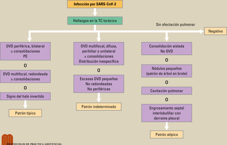
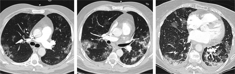
Chest x-ray and computed tomography (CT) scans are important pillars for the diagnosis of lung involvement in COVID-19. The radiological image is typically characterized by peripheral, bilateral ground glass opacities (GGO), mainly located in the lower lobes. The limited sensitivity and specificity of these imaging techniques and possible atypical morphological or topographical presentations make it necessary to always rule out other infectious and non-infectious diseases. Therefore, it is fundamental to consider the patient's clinical and analytical data and the epidemiological circumstances.

摘要

胸部X光和计算机断层扫描(CT)是诊断COVID-19肺部受累情况的重要支柱。放射影像的典型特征是外周双侧磨玻璃影(GGO),主要位于下叶。这些成像技术的敏感性和特异性有限,以及可能出现的非典型形态或部位表现,使得始终有必要排除其他感染性和非感染性疾病。因此,综合考虑患者的临床和分析数据以及流行病学情况至关重要。

https://cdn.ncbi.nlm.nih.gov/pmc/blobs/d5d3/9098090/35b3ff181e42/gr2_lrg.jpg
https://cdn.ncbi.nlm.nih.gov/pmc/blobs/d5d3/9098090/ffce2dbc0da3/gr1_lrg.jpg
https://cdn.ncbi.nlm.nih.gov/pmc/blobs/d5d3/9098090/35b3ff181e42/gr2_lrg.jpg
https://cdn.ncbi.nlm.nih.gov/pmc/blobs/d5d3/9098090/ffce2dbc0da3/gr1_lrg.jpg
https://cdn.ncbi.nlm.nih.gov/pmc/blobs/d5d3/9098090/35b3ff181e42/gr2_lrg.jpg

相似文献

1
[Diagnostic protocol for febrile lung infiltrates during the COVID-19 pandemic].[新型冠状病毒肺炎疫情期间发热性肺部浸润的诊断方案]
Medicine (Madr). 2022 May;13(55):3261-3265. doi: 10.1016/j.med.2022.05.009. Epub 2022 May 12.
2
Comparison of the computed tomography findings in COVID-19 and other viral pneumonia in immunocompetent adults: a systematic review and meta-analysis.COVID-19 与免疫功能正常成年人其他病毒性肺炎的计算机断层扫描表现比较:系统评价和荟萃分析。
Eur Radiol. 2020 Dec;30(12):6485-6496. doi: 10.1007/s00330-020-07018-x. Epub 2020 Jun 27.
3
Ground-glass opacity (GGO): a review of the differential diagnosis in the era of COVID-19.磨玻璃密度影(GGO):COVID-19 时代的鉴别诊断综述。
Jpn J Radiol. 2021 Aug;39(8):721-732. doi: 10.1007/s11604-021-01120-w. Epub 2021 Apr 26.
4
Imaging and clinical features of patients with 2019 novel coronavirus SARS-CoV-2.新型冠状病毒 2019 年 SARS-CoV-2 患者的影像学和临床特征。
Eur J Nucl Med Mol Imaging. 2020 May;47(5):1275-1280. doi: 10.1007/s00259-020-04735-9. Epub 2020 Feb 28.
5
Lung perfusion with dual-energy multi-detector row CT: can it help recognize ground glass opacities of vascular origin?双能多排 CT 肺灌注:有助于识别血管源性磨玻璃影吗?
Acad Radiol. 2010 May;17(5):587-94. doi: 10.1016/j.acra.2009.12.013. Epub 2010 Mar 3.
6
CT-scan findings of COVID-19 pneumonia based on the time elapsed from the beginning of symptoms to the CT imaging evaluation: a descriptive study in Iran.基于从症状出现到 CT 影像评估时间的 COVID-19 肺炎 CT 扫描结果:伊朗的一项描述性研究。
Rom J Intern Med. 2020 Dec 17;58(4):242-250. doi: 10.2478/rjim-2020-0019. Print 2020 Dec 1.
7
COVID-19 pneumonia: CT findings of 122 patients and differentiation from influenza pneumonia.新型冠状病毒肺炎:122例患者的CT表现及与流感肺炎的鉴别诊断
Eur Radiol. 2020 Oct;30(10):5463-5469. doi: 10.1007/s00330-020-06928-0. Epub 2020 May 12.
8
Differential diagnosis of coronavirus disease 2019 from community-acquired-pneumonia by computed tomography scan and follow-up.通过计算机断层扫描和随访对 2019 年冠状病毒病与社区获得性肺炎进行鉴别诊断。
Infect Dis Poverty. 2020 Aug 26;9(1):118. doi: 10.1186/s40249-020-00737-9.
9
[Spatial and temporal distribution and predictive value of chest CT scoring in patients with COVID-19].新型冠状病毒肺炎患者胸部CT评分的时空分布及预测价值
Zhonghua Jie He He Hu Xi Za Zhi. 2021 Mar 12;44(3):230-236. doi: 10.3760/cma.j.cn112147-20200522-00626.
10
Atypical Chest Computed Tomography Finding of Predominant Interstitial Thickening in a Patient with Coronavirus Disease 2019 (COVID-19) Pneumonia.一名2019冠状病毒病(COVID-19)肺炎患者胸部计算机断层扫描的非典型表现:以间质增厚为主
Am J Case Rep. 2020 Sep 21;21:e926781. doi: 10.12659/AJCR.926781.

本文引用的文献

1
Ground-glass opacity (GGO): a review of the differential diagnosis in the era of COVID-19.磨玻璃密度影(GGO):COVID-19 时代的鉴别诊断综述。
Jpn J Radiol. 2021 Aug;39(8):721-732. doi: 10.1007/s11604-021-01120-w. Epub 2021 Apr 26.
2
Radiologic diagnosis of patients with COVID-19.COVID-19 患者的放射学诊断。
Radiologia (Engl Ed). 2021 Jan-Feb;63(1):56-73. doi: 10.1016/j.rx.2020.11.001. Epub 2020 Nov 24.
3
Thoracic tomographic manifestations in symptomatic respiratory patients with COVID-19.有症状的COVID-19呼吸道患者的胸部断层扫描表现。
Radiol Bras. 2020 Jul-Aug;53(4):255-261. doi: 10.1590/0100-3984.2020.0030.
4
Radiological Society of North America Expert Consensus Statement on Reporting Chest CT Findings Related to COVID-19. Endorsed by the Society of Thoracic Radiology, the American College of Radiology, and RSNA - Secondary Publication.北美放射学会关于报告与 COVID-19 相关的胸部 CT 结果的专家共识声明。得到胸放射学会、美国放射学会和 RSNA 的认可 - 二次出版物。
J Thorac Imaging. 2020 Jul;35(4):219-227. doi: 10.1097/RTI.0000000000000524.
5
The Role of Chest Imaging in Patient Management during the COVID-19 Pandemic: A Multinational Consensus Statement from the Fleischner Society.《COVID-19 大流行期间患者管理中的胸部影像学作用:Fleischner 学会的一项多国共识声明》。
Radiology. 2020 Jul;296(1):172-180. doi: 10.1148/radiol.2020201365. Epub 2020 Apr 7.
6
Frequency and Distribution of Chest Radiographic Findings in Patients Positive for COVID-19.COVID-19 阳性患者的胸部 X 线表现的频率和分布。
Radiology. 2020 Aug;296(2):E72-E78. doi: 10.1148/radiol.2020201160. Epub 2020 Mar 27.
7
Performance of Radiologists in Differentiating COVID-19 from Non-COVID-19 Viral Pneumonia at Chest CT.放射科医生在胸部 CT 鉴别 COVID-19 与非 COVID-19 病毒性肺炎中的表现。
Radiology. 2020 Aug;296(2):E46-E54. doi: 10.1148/radiol.2020200823. Epub 2020 Mar 10.
8
An official American Thoracic Society/European Respiratory Society statement: Update of the international multidisciplinary classification of the idiopathic interstitial pneumonias.美国胸科学会/欧洲呼吸学会官方声明:特发性间质性肺炎的国际多学科分类的更新。
Am J Respir Crit Care Med. 2013 Sep 15;188(6):733-48. doi: 10.1164/rccm.201308-1483ST.
9
Acute pulmonary injury: high-resolution CT and histopathological spectrum.急性肺损伤:高分辨率 CT 与组织病理学表现。
Br J Radiol. 2013 Jul;86(1027):20120614. doi: 10.1259/bjr.20120614. Epub 2013 May 9.
10
Does this dyspneic patient in the emergency department have congestive heart failure?这位在急诊科出现呼吸困难的患者是否患有充血性心力衰竭?
JAMA. 2005 Oct 19;294(15):1944-56. doi: 10.1001/jama.294.15.1944.