Robarts Research Institute, Western University, London, Ontario, Canada.
Department of Medical Biophysics, Western University, London, Ontario, Canada.
BMJ Open Respir Res. 2022 May;9(1). doi: 10.1136/bmjresp-2022-001235.
Patients often report persistent symptoms beyond the acute infectious phase of COVID-19. Hyperpolarised Xe MRI provides a way to directly measure airway functional abnormalities; the clinical relevance of Xe MRI ventilation defects in ever-hospitalised and never-hospitalised patients who had COVID-19 has not been ascertained. It remains unclear if persistent symptoms beyond the infectious phase are related to small airways disease and ventilation heterogeneity. Hence, we measured Xe MRI ventilation defects, pulmonary function and symptoms in ever-hospitalised and never-hospitalised patients who had COVID-19 with persistent symptoms consistent with post-acute COVID-19 syndrome (PACS).
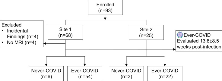
Consenting participants with a confirmed diagnosis of PACS completed Xe MRI, CT, spirometry, multi-breath inert-gas washout, 6-minute walk test, St. George's Respiratory Questionnaire (SGRQ), modified Medical Research Council (mMRC) dyspnoea scale, modified Borg scale and International Physical Activity Questionnaire. Consenting ever-COVID volunteers completed Xe MRI and pulmonary function tests only.
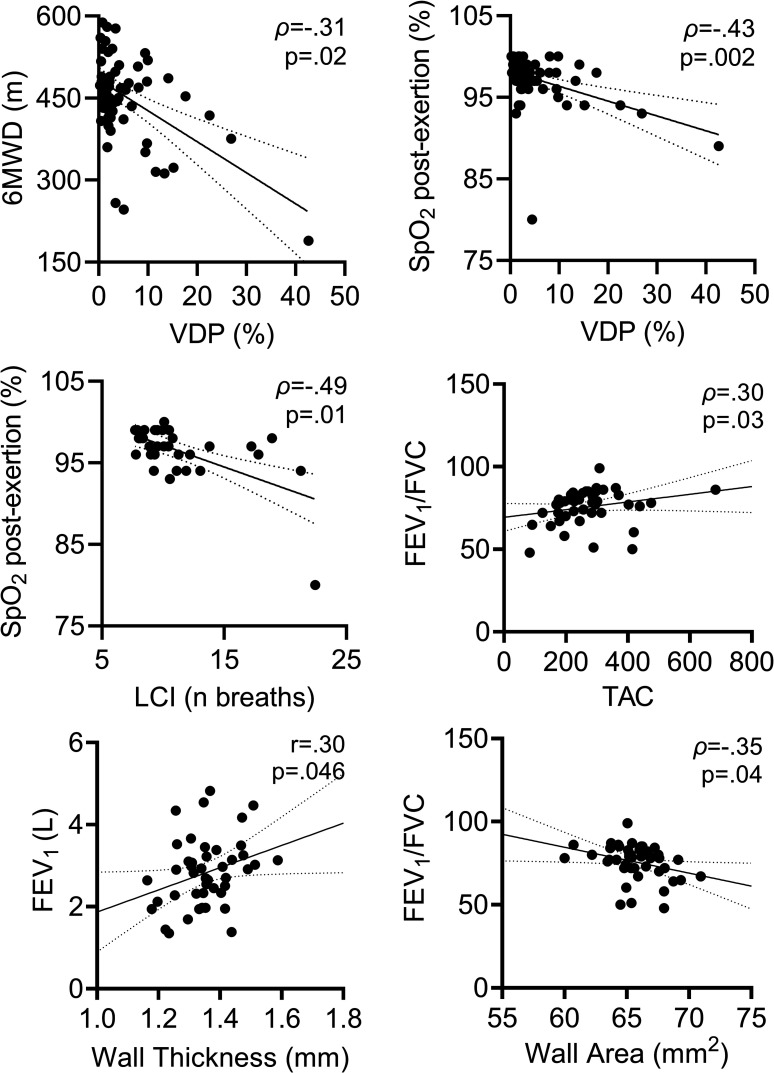
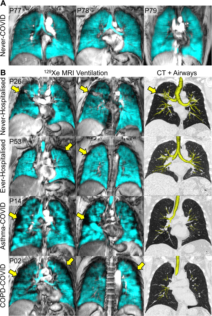
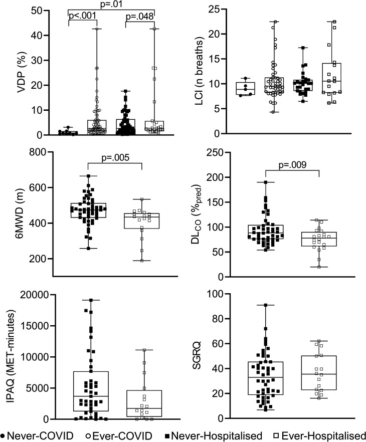
Seventy-six post-COVID and nine never-COVID participants were evaluated. Ventilation defect per cent (VDP) was abnormal and significantly greater in ever-COVID as compared with never-COVID participants (p<0.001) and significantly greater in ever-hospitalised compared with never-hospitalised participants who had COVID-19 (p=0.048), in whom diffusing capacity of the lung for carbon-monoxide (p=0.009) and 6-minute walk distance (6MWD) (p=0.005) were also significantly different. Xe MRI VDP was also related to the 6MWD (p=0.02) and post-exertional SpO (p=0.002). Participants with abnormal VDP (≥4.3%) had significantly worse 6MWD (p=0.003) and post-exertional SpO (p=0.03).
Xe MRI VDP was significantly worse in ever-hospitalised as compared with never-hospitalised participants and was related to 6MWD and exertional SpO but not SGRQ or mMRC scores.
NCT05014516.
患者在 COVID-19 的急性感染期过后常常报告持续存在的症状。超极化氙气 MRI 提供了一种直接测量气道功能异常的方法;在因 COVID-19 而住院和未住院的患者中,Xe MRI 通气缺陷的临床相关性尚未确定。目前尚不清楚感染期过后的持续症状是否与小气道疾病和通气异质性有关。因此,我们在符合急性 COVID-19 后综合征 (PACS) 标准的因 COVID-19 而持续存在症状的住院和未住院患者中测量了 Xe MRI 通气缺陷、肺功能和症状。
同意参加研究的 PACS 确诊患者完成了 Xe MRI、CT、肺量计检查、多呼吸惰性气体洗脱、6 分钟步行试验、圣乔治呼吸问卷 (SGRQ)、改良版医学研究委员会呼吸困难量表、改良版 Borg 量表和国际体力活动问卷。同意的曾 COVID 志愿者仅完成了 Xe MRI 和肺功能测试。
共评估了 76 名新冠后患者和 9 名从未感染过 COVID-19 的患者。与从未感染过 COVID-19 的患者相比,Xe MRI 通气缺陷百分比(VDP)在曾 COVID 患者中异常且显著更高(p<0.001),且在因 COVID-19 而住院的患者中显著更高(p=0.048),在这些患者中,一氧化碳弥散量(p=0.009)和 6 分钟步行距离(6MWD)(p=0.005)也有显著差异。Xe MRI VDP 还与 6MWD(p=0.02)和运动后 SpO(p=0.002)相关。VDP 异常(≥4.3%)的患者 6MWD 明显更差(p=0.003),运动后 SpO 明显更低(p=0.03)。
与未住院的患者相比,曾住院的患者 Xe MRI VDP 明显更差,且与 6MWD 和运动后 SpO 相关,但与 SGRQ 或 mMRC 评分无关。
NCT05014516。