• 文献检索
  • 文档翻译
  • 深度研究
  • 学术资讯
  • Suppr Zotero 插件Zotero 插件
  • 邀请有礼
  • 套餐&价格
  • 历史记录
应用&插件
Suppr Zotero 插件Zotero 插件浏览器插件Mac 客户端Windows 客户端微信小程序
定价
高级版会员购买积分包购买API积分包
服务
文献检索文档翻译深度研究API 文档MCP 服务
关于我们
关于 Suppr公司介绍联系我们用户协议隐私条款
关注我们

Suppr 超能文献

核心技术专利:CN118964589B侵权必究
粤ICP备2023148730 号-1Suppr @ 2026

文献检索

告别复杂PubMed语法,用中文像聊天一样搜索,搜遍4000万医学文献。AI智能推荐,让科研检索更轻松。

立即免费搜索

文件翻译

保留排版,准确专业,支持PDF/Word/PPT等文件格式,支持 12+语言互译。

免费翻译文档

深度研究

AI帮你快速写综述,25分钟生成高质量综述,智能提取关键信息,辅助科研写作。

立即免费体验

单细胞转录组揭示了斑马鱼卵巢发育和功能的见解。

Single-cell transcriptome reveals insights into the development and function of the zebrafish ovary.

机构信息

Department of Molecular and Cellular Biology, University of California, Davis, Davis, United States.

Mount Desert Island Biological Laboratory, Bar Harbor, United States.

出版信息

Elife. 2022 May 19;11:e76014. doi: 10.7554/eLife.76014.

DOI:10.7554/eLife.76014
PMID:35588359
原文链接:https://pmc.ncbi.nlm.nih.gov/articles/PMC9191896/
Abstract

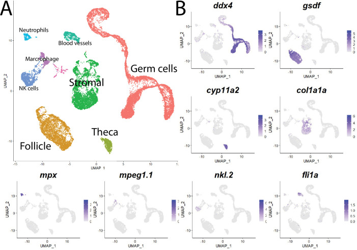
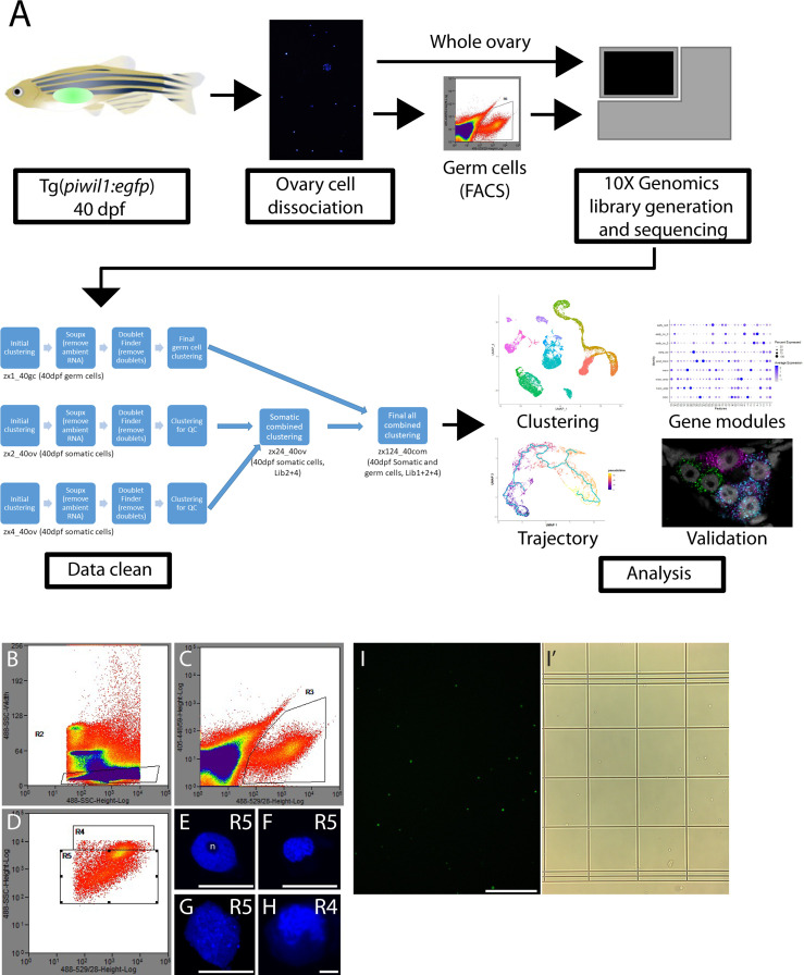
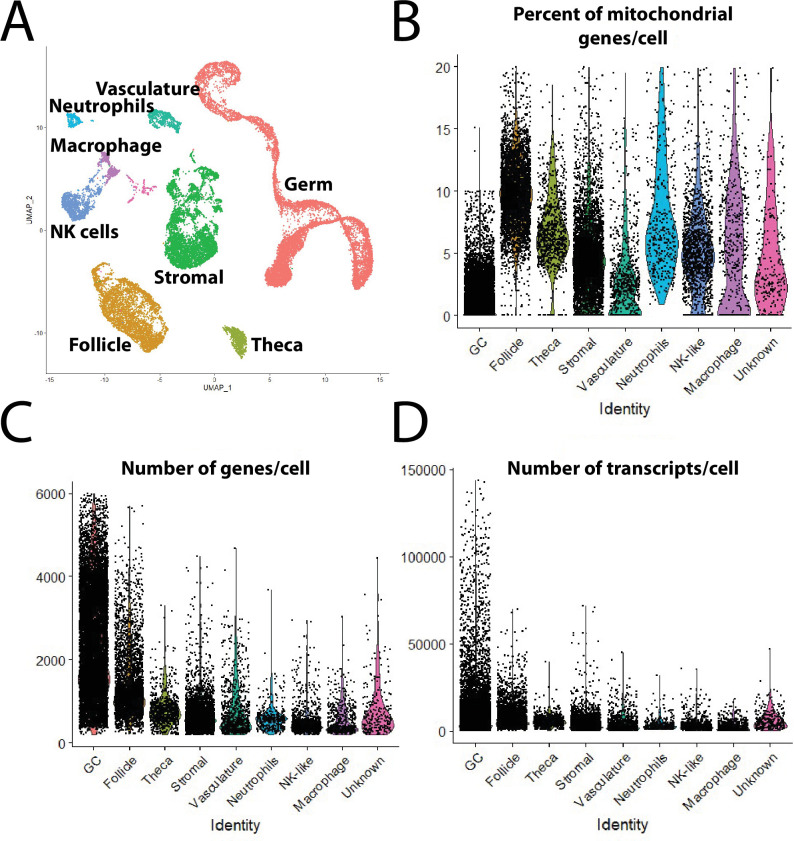
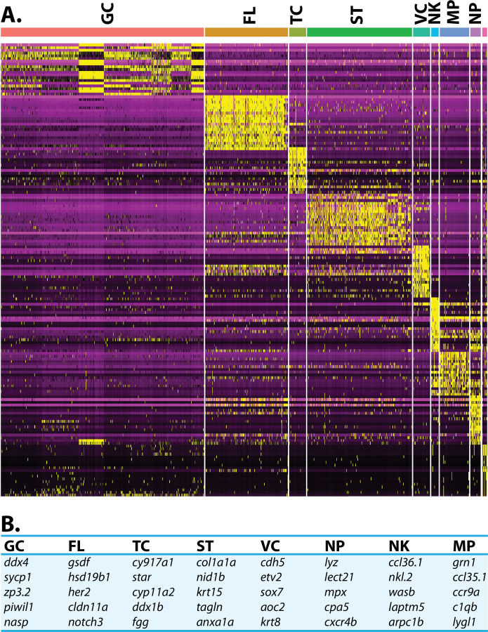
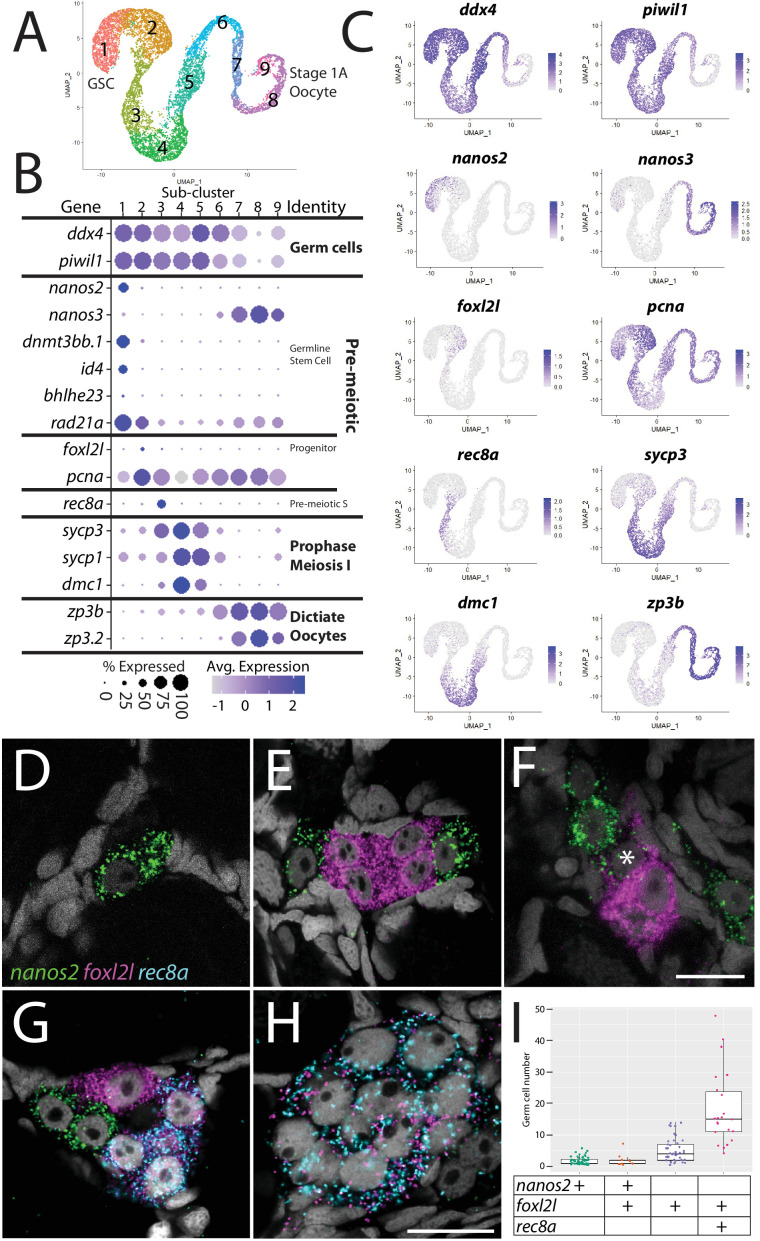
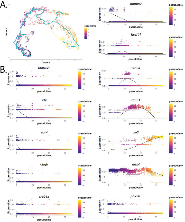
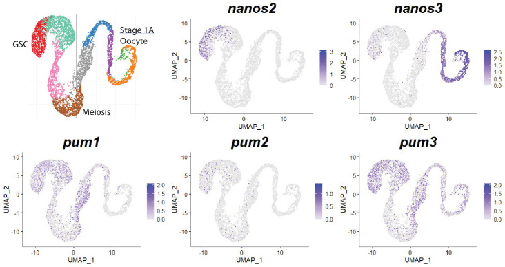
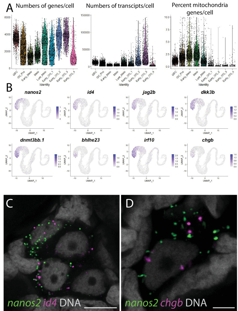
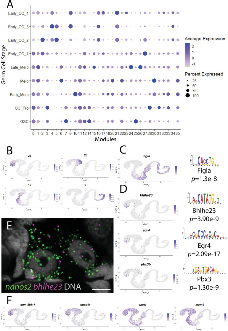
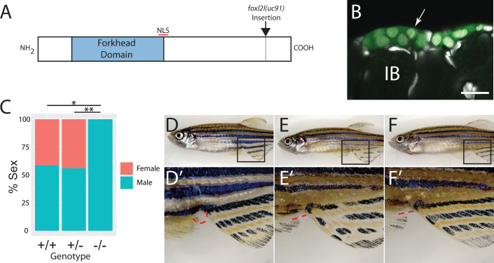
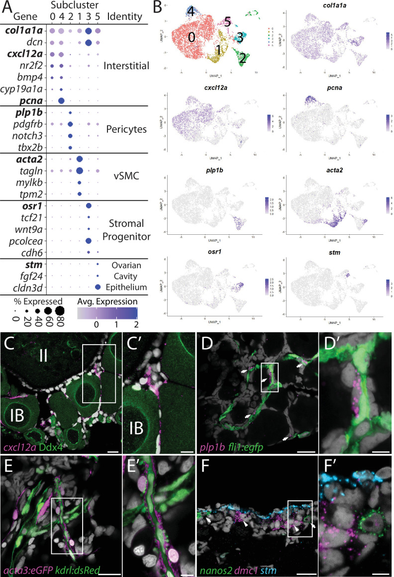
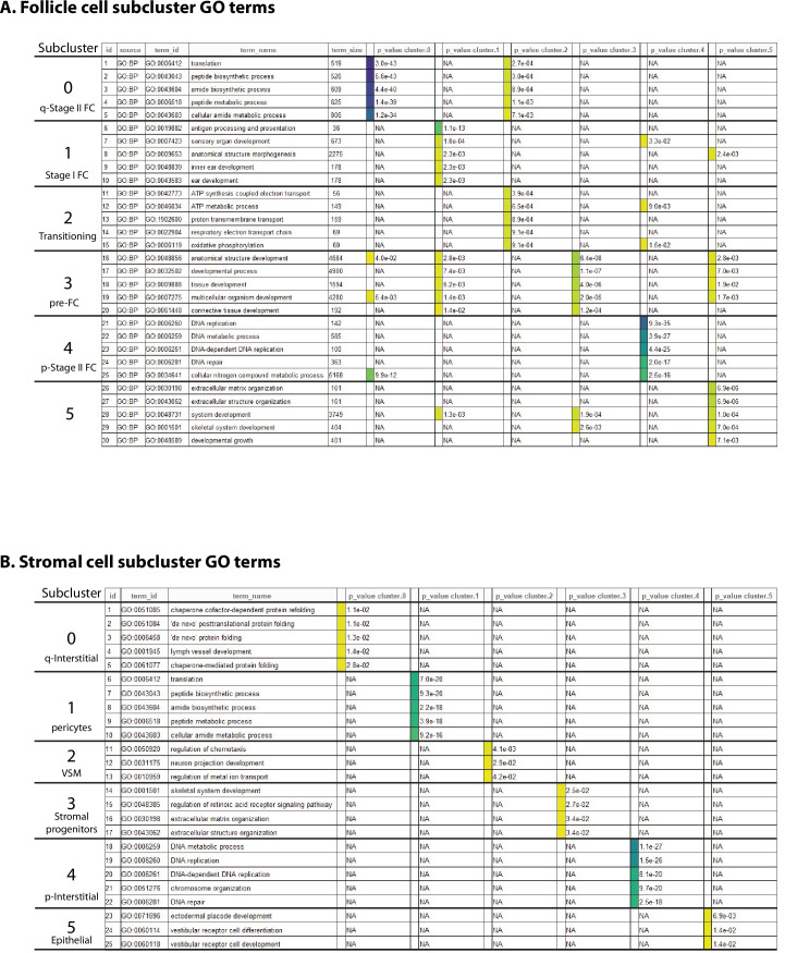
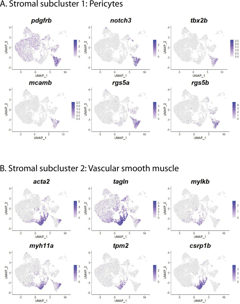
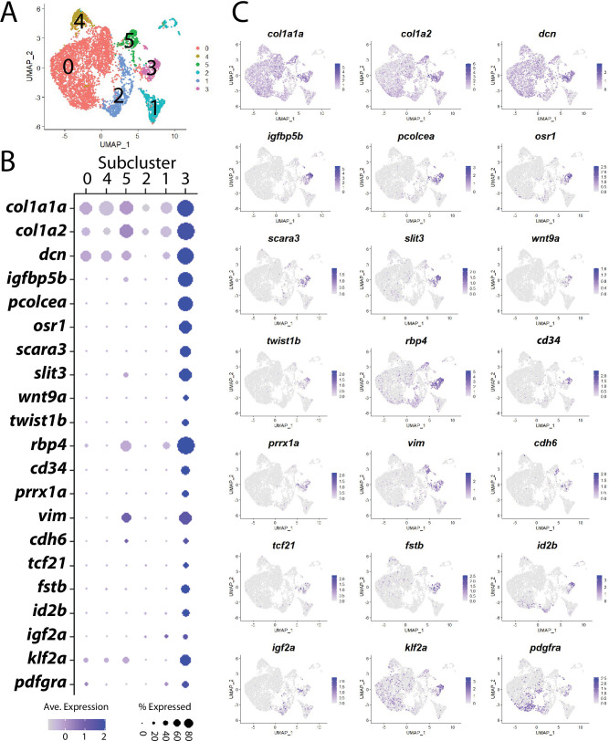
Zebrafish are an established research organism that has made many contributions to our understanding of vertebrate tissue and organ development, yet there are still significant gaps in our understanding of the genes that regulate gonad development, sex, and reproduction. Unlike the development of many organs, such as the brain and heart that form during the first few days of development, zebrafish gonads do not begin to form until the larval stage (≥5 days post-fertilization). Thus, forward genetic screens have identified very few genes required for gonad development. In addition, bulk RNA-sequencing studies that identify genes expressed in the gonads do not have the resolution necessary to define minor cell populations that may play significant roles in the development and function of these organs. To overcome these limitations, we have used single-cell RNA sequencing to determine the transcriptomes of cells isolated from juvenile zebrafish ovaries. This resulted in the profiles of 10,658 germ cells and 14,431 somatic cells. Our germ cell data represents all developmental stages from germline stem cells to early meiotic oocytes. Our somatic cell data represents all known somatic cell types, including follicle cells, theca cells, and ovarian stromal cells. Further analysis revealed an unexpected number of cell subpopulations within these broadly defined cell types. To further define their functional significance, we determined the location of these cell subpopulations within the ovary. Finally, we used gene knockout experiments to determine the roles of and for oocyte development and sex determination and/or differentiation, respectively. Our results reveal novel insights into zebrafish ovarian development and function, and the transcriptome profiles will provide a valuable resource for future studies.

摘要

斑马鱼是一种成熟的研究生物,它为我们理解脊椎动物组织和器官的发育做出了许多贡献,但我们对调节性腺发育、性别和生殖的基因仍知之甚少。与大脑和心脏等许多器官在发育的头几天形成不同,斑马鱼的性腺直到幼体期(受精后≥5 天)才开始形成。因此,正向遗传筛选只鉴定出了很少的性腺发育所必需的基因。此外,用于鉴定在性腺中表达的基因的批量 RNA 测序研究,没有确定可能在这些器官的发育和功能中发挥重要作用的少数细胞群体的分辨率。为了克服这些限制,我们使用单细胞 RNA 测序来确定从小型斑马鱼卵巢中分离出的细胞的转录组。这导致了 10658 个生殖细胞和 14431 个体细胞的图谱。我们的生殖细胞数据代表了从生殖干细胞到早期减数分裂卵母细胞的所有发育阶段。我们的体细胞数据代表了所有已知的体细胞类型,包括滤泡细胞、膜细胞和卵巢基质细胞。进一步的分析揭示了这些广泛定义的细胞类型中存在大量的细胞亚群。为了进一步确定它们的功能意义,我们确定了这些细胞亚群在卵巢中的位置。最后,我们使用基因敲除实验确定了和分别在卵母细胞发育和性别决定/分化中的作用。我们的结果揭示了斑马鱼卵巢发育和功能的新见解,转录组图谱将为未来的研究提供有价值的资源。

https://cdn.ncbi.nlm.nih.gov/pmc/blobs/9f0d/9191896/48e5fde5774d/elife-76014-fig9-figsupp1.jpg
https://cdn.ncbi.nlm.nih.gov/pmc/blobs/9f0d/9191896/9b1b29a4709a/elife-76014-fig1.jpg
https://cdn.ncbi.nlm.nih.gov/pmc/blobs/9f0d/9191896/600b57d98a15/elife-76014-fig1-figsupp1.jpg
https://cdn.ncbi.nlm.nih.gov/pmc/blobs/9f0d/9191896/06d60d059089/elife-76014-fig1-figsupp2.jpg
https://cdn.ncbi.nlm.nih.gov/pmc/blobs/9f0d/9191896/054ba9822f24/elife-76014-fig1-figsupp3.jpg
https://cdn.ncbi.nlm.nih.gov/pmc/blobs/9f0d/9191896/0cd9a5bcbf9a/elife-76014-fig2.jpg
https://cdn.ncbi.nlm.nih.gov/pmc/blobs/9f0d/9191896/b9383ef8a66f/elife-76014-fig2-figsupp1.jpg
https://cdn.ncbi.nlm.nih.gov/pmc/blobs/9f0d/9191896/bb15635fd4ed/elife-76014-fig2-figsupp2.jpg
https://cdn.ncbi.nlm.nih.gov/pmc/blobs/9f0d/9191896/7047b5fca588/elife-76014-fig2-figsupp3.jpg
https://cdn.ncbi.nlm.nih.gov/pmc/blobs/9f0d/9191896/816e6c77f625/elife-76014-fig2-figsupp4.jpg
https://cdn.ncbi.nlm.nih.gov/pmc/blobs/9f0d/9191896/d90decddc9c5/elife-76014-fig2-figsupp5.jpg
https://cdn.ncbi.nlm.nih.gov/pmc/blobs/9f0d/9191896/5eb27d5812a4/elife-76014-fig3.jpg
https://cdn.ncbi.nlm.nih.gov/pmc/blobs/9f0d/9191896/c8a88ee9c158/elife-76014-fig4.jpg
https://cdn.ncbi.nlm.nih.gov/pmc/blobs/9f0d/9191896/5bd26d8db434/elife-76014-fig4-figsupp1.jpg
https://cdn.ncbi.nlm.nih.gov/pmc/blobs/9f0d/9191896/1eb4c6e7218b/elife-76014-fig4-figsupp2.jpg
https://cdn.ncbi.nlm.nih.gov/pmc/blobs/9f0d/9191896/8c8a5756a6d9/elife-76014-fig4-figsupp3.jpg
https://cdn.ncbi.nlm.nih.gov/pmc/blobs/9f0d/9191896/a3851feba6d2/elife-76014-fig5.jpg
https://cdn.ncbi.nlm.nih.gov/pmc/blobs/9f0d/9191896/29ea389b4c9b/elife-76014-fig5-figsupp1.jpg
https://cdn.ncbi.nlm.nih.gov/pmc/blobs/9f0d/9191896/680104524071/elife-76014-fig6.jpg
https://cdn.ncbi.nlm.nih.gov/pmc/blobs/9f0d/9191896/edb15252bbb3/elife-76014-fig6-figsupp1.jpg
https://cdn.ncbi.nlm.nih.gov/pmc/blobs/9f0d/9191896/34f68efcf572/elife-76014-fig6-figsupp2.jpg
https://cdn.ncbi.nlm.nih.gov/pmc/blobs/9f0d/9191896/71de0aed1757/elife-76014-fig6-figsupp3.jpg
https://cdn.ncbi.nlm.nih.gov/pmc/blobs/9f0d/9191896/1b0dad2eae6c/elife-76014-fig6-figsupp4.jpg
https://cdn.ncbi.nlm.nih.gov/pmc/blobs/9f0d/9191896/340882c0db4b/elife-76014-fig7.jpg
https://cdn.ncbi.nlm.nih.gov/pmc/blobs/9f0d/9191896/4a0974076a60/elife-76014-fig8.jpg
https://cdn.ncbi.nlm.nih.gov/pmc/blobs/9f0d/9191896/577c727eb205/elife-76014-fig8-figsupp1.jpg
https://cdn.ncbi.nlm.nih.gov/pmc/blobs/9f0d/9191896/aa7eabb86e6f/elife-76014-fig9.jpg
https://cdn.ncbi.nlm.nih.gov/pmc/blobs/9f0d/9191896/48e5fde5774d/elife-76014-fig9-figsupp1.jpg

相似文献

1
Single-cell transcriptome reveals insights into the development and function of the zebrafish ovary.单细胞转录组揭示了斑马鱼卵巢发育和功能的见解。
Elife. 2022 May 19;11:e76014. doi: 10.7554/eLife.76014.
2
Erratum: Eyestalk Ablation to Increase Ovarian Maturation in Mud Crabs.勘误:切除眼柄以增加泥蟹的卵巢成熟度。
J Vis Exp. 2023 May 26(195). doi: 10.3791/6561.
3
Fibroblast growth factor signaling is required for early somatic gonad development in zebrafish.成纤维细胞生长因子信号传导是斑马鱼早期体细胞性腺发育所必需的。
PLoS Genet. 2017 Sep 5;13(9):e1006993. doi: 10.1371/journal.pgen.1006993. eCollection 2017 Sep.
4
Dynamics of the transcriptional landscape during human fetal testis and ovary development.人类胎儿睾丸和卵巢发育过程中转录组景观的动态变化。
Hum Reprod. 2020 May 1;35(5):1099-1119. doi: 10.1093/humrep/deaa041.
5
Changes in the morphology and gene expression of developing zebrafish gonads.发育中的斑马鱼性腺的形态学和基因表达变化。
Gen Comp Endocrinol. 2018 Sep 1;265:154-159. doi: 10.1016/j.ygcen.2018.01.026. Epub 2018 Jan 31.
6
is a germ cell-intrinsic gatekeeper of oogenesis in zebrafish.它是斑马鱼卵子发生过程中生殖细胞内的一个关键调控因子。
Zool Res. 2024 Sep 18;45(5):1116-1130. doi: 10.24272/j.issn.2095-8137.2024.046.
7
Transcriptome profiling reveals male- and female-specific gene expression pattern and novel gene candidates for the control of sex determination and gonad development in Xenopus laevis.转录组谱分析揭示了非洲爪蟾性别决定和性腺发育的性别特异性基因表达模式和新的候选基因。
Dev Genes Evol. 2019 May;229(2-3):53-72. doi: 10.1007/s00427-019-00630-y. Epub 2019 Apr 10.
8
Transcriptional profiling validates involvement of extracellular matrix and proteinases genes in mouse gonad development.转录谱分析验证了细胞外基质和蛋白酶基因在小鼠性腺发育中的作用。
Mech Dev. 2018 Feb;149:9-19. doi: 10.1016/j.mod.2017.11.001. Epub 2017 Nov 9.
9
Direct male development in chromosomally ZZ zebrafish.染色体为ZZ的斑马鱼的雄性直接发育。
Front Cell Dev Biol. 2024 Mar 11;12:1362228. doi: 10.3389/fcell.2024.1362228. eCollection 2024.
10
Retinoic acid metabolic genes, meiosis, and gonadal sex differentiation in zebrafish.视黄酸代谢基因、减数分裂和斑马鱼的性腺性别分化。
PLoS One. 2013 Sep 10;8(9):e73951. doi: 10.1371/journal.pone.0073951. eCollection 2013.

引用本文的文献

1
Distinct cellular and reproductive consequences of meiotic chromosome synapsis defects in syce2 and sycp1 mutant zebrafish.syce2和sycp1突变斑马鱼减数分裂染色体联会缺陷的不同细胞和生殖后果
PLoS Genet. 2025 Sep 5;21(9):e1011656. doi: 10.1371/journal.pgen.1011656. eCollection 2025 Sep.
2
Single-Nucleus Transcriptome Sequencing Unravels Physiological Differences in Holstein Cows Under Different Physiological States.单核转录组测序揭示不同生理状态下荷斯坦奶牛的生理差异
Genes (Basel). 2025 Aug 3;16(8):931. doi: 10.3390/genes16080931.
3
The Role of Neurohypophysial Hormones in the Endocrine and Paracrine Control of Gametogenesis in Fish.

本文引用的文献

1
TCF21 mesenchymal cells contribute to testis somatic cell development, homeostasis, and regeneration in mice.TCF21 间质细胞有助于小鼠睾丸体细胞的发育、稳态和再生。
Nat Commun. 2021 Jun 23;12(1):3876. doi: 10.1038/s41467-021-24130-8.
2
Histological Analysis of Gonads in Zebrafish.斑马鱼性腺的组织学分析。
Methods Mol Biol. 2021;2218:253-263. doi: 10.1007/978-1-0716-0970-5_20.
3
SoupX removes ambient RNA contamination from droplet-based single-cell RNA sequencing data.SoupX 去除了基于液滴的单细胞 RNA 测序数据中的环境 RNA 污染。
神经垂体激素在鱼类配子发生的内分泌和旁分泌控制中的作用
Cells. 2025 Jul 10;14(14):1061. doi: 10.3390/cells14141061.
4
Clonal analysis reveals differential PGC contributions to the early germline and ovarian reserve.克隆分析揭示了原始生殖细胞对早期生殖系和卵巢储备的不同贡献。
bioRxiv. 2025 May 7:2025.04.30.651497. doi: 10.1101/2025.04.30.651497.
5
Integrative single-cell transcriptomics of sheep ovarian development reveals dynamic transcriptional programs relevant to reproductive traits.绵羊卵巢发育的整合单细胞转录组学揭示了与繁殖性状相关的动态转录程序。
iScience. 2025 Apr 14;28(5):112422. doi: 10.1016/j.isci.2025.112422. eCollection 2025 May 16.
6
Integration of hyperspectral imaging and transcriptomics from individual cells with SpectralSeq.利用SpectralSeq将高光谱成像与单个细胞的转录组学相结合。
Genome Res. 2025 Aug 1;35(8):1809-1820. doi: 10.1101/gr.280014.124.
7
Single-cell transcriptomes of zebrafish germline reveal progenitor types and feminization by Foxl2l.斑马鱼生殖系的单细胞转录组揭示了祖细胞类型以及Foxl2l诱导的雌性化现象。
Elife. 2025 Jun 11;14:e100204. doi: 10.7554/eLife.100204.
8
Differential expression of sex regulatory genes in gonads of Astyanax mexicanus surface fish and cavefish.墨西哥丽脂鲤表层鱼和洞穴鱼性腺中性别调控基因的差异表达。
BMC Ecol Evol. 2025 May 28;25(1):57. doi: 10.1186/s12862-025-02376-w.
9
Folliculogenesis and follicular growth in the endangered fish Brycon opalinus, with emphasis on ovarian matrix remodeling.濒危鱼类欧帕林脂鲤的卵泡发生和卵泡生长,重点关注卵巢基质重塑。
Cell Tissue Res. 2025 May 19. doi: 10.1007/s00441-025-03982-2.
10
Reproductive adaptation of Astyanax mexicanus under nutrient limitation.墨西哥丽脂鲤在营养限制下的生殖适应性
Dev Biol. 2025 Jul;523:82-98. doi: 10.1016/j.ydbio.2025.04.006. Epub 2025 Apr 11.
Gigascience. 2020 Dec 26;9(12). doi: 10.1093/gigascience/giaa151.
4
Transcriptional landscape of the embryonic chicken Müllerian duct.鸡胚缪勒氏管的转录组图谱。
BMC Genomics. 2020 Oct 2;21(1):688. doi: 10.1186/s12864-020-07106-8.
5
The ovarian stroma as a new frontier.卵巢间质:新前沿
Reproduction. 2020 Sep;160(3):R25-R39. doi: 10.1530/REP-19-0501.
6
Efficient targeted integration directed by short homology in zebrafish and mammalian cells.通过短同源序列在斑马鱼和哺乳动物细胞中的高效靶向整合。
Elife. 2020 May 15;9:e53968. doi: 10.7554/eLife.53968.
7
, a sexual switch in germ cells, initiates two independent molecular pathways for commitment to oogenesis in medaka.在鱼类中,生殖细胞的性转换启动了两条独立的分子途径,使生殖细胞向卵母细胞分化。
Proc Natl Acad Sci U S A. 2020 Jun 2;117(22):12174-12181. doi: 10.1073/pnas.1918556117. Epub 2020 May 14.
8
Phylogeny of teleost connexins reveals highly inconsistent intra- and interspecies use of nomenclature and misassemblies in recent teleost chromosome assemblies.鱼类连接蛋白的系统发育揭示了近期鱼类染色体组装中高度不一致的种内和种间命名法使用以及错误组装。
BMC Genomics. 2020 Mar 11;21(1):223. doi: 10.1186/s12864-020-6620-2.
9
Single-cell analysis of human ovarian cortex identifies distinct cell populations but no oogonial stem cells.单细胞分析人类卵巢皮质可鉴定出不同的细胞群体,但不存在卵原干细胞。
Nat Commun. 2020 Mar 2;11(1):1147. doi: 10.1038/s41467-020-14936-3.
10
Endocrine and local signaling interact to regulate spermatogenesis in zebrafish: follicle-stimulating hormone, retinoic acid and androgens.内分泌和局部信号相互作用调节斑马鱼的精子发生:促卵泡激素、视黄酸和雄激素。
Development. 2019 Nov 4;146(21):dev178665. doi: 10.1242/dev.178665.