Yuan Shiting, Wu Huiqin, Wu Yun, Xu Huazhen, Yu Jianping, Zhong Yuan, Zhang Ning, Li Jinyang, Xu Qianwen, Wang Chun
Nanjing Brain Hospital Affiliated to Nanjing Medical University, Nanjing, China.
School of Psychology, Nanjing Normal University, Nanjing, China.
Front Psychol. 2022 May 3;13:853804. doi: 10.3389/fpsyg.2022.853804. eCollection 2022.
Cognitive behavioral therapy (CBT) is a first-line psychotherapeutic treatment that has been recommended for psychiatric disorders. Prior neuroimaging studies have provided preliminary evidence suggesting that CBT can have an impact on the activity of brain regions and functional integration between regions. However, the results are far from conclusive. The present article aimed to detect characteristic changes in brain activation following CBT across psychiatric disorders.
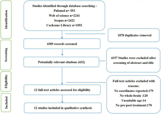
Web of Science, Cochrane Library, Scopus, and PubMed databases were searched to identify whole-brain functional neuroimaging studies of CBT through 4 August 2021. To be included in the meta-analysis, studies were required to examine functional activation changes between pre-and post-CBT. The included studies were then divided into subgroups according to different task paradigms. Then, an activation likelihood estimation algorithm (ALE) was performed in the different meta-analyses to identify whether brain regions showed consistent effects. Finally, brain regions identified from the meta-analysis were categorized into eight functional networks according to the spatial correlation values between independent components and the template.
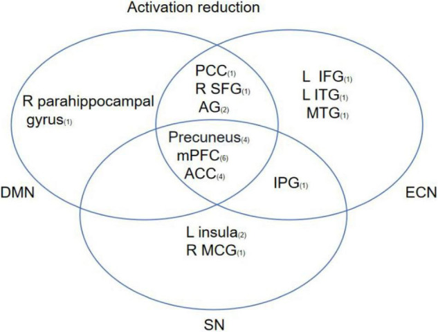
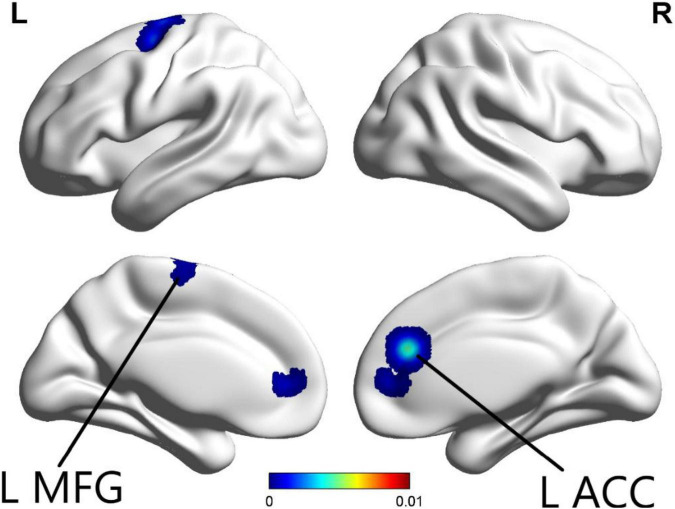
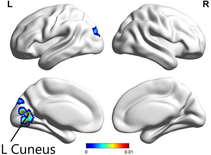
In total, 13 studies met inclusion criteria. Three different meta-analyses were performed separately for total tasks, emotion tasks, and cognition tasks. In the total task ALE meta-analysis, the left precuneus was found to have decreased activation. For the cognition task ALE meta-analysis, left anterior cingulate (ACC) and left middle frontal gyrus (MFG) were found to have decreased activation following CBT. However, the emotion task ALE meta-analysis did not find any specific brain regions showing consistent effects. A review of included studies revealed default mode network (DMN), executive control network (ECN), and salience network (SN) were the most relevant among the eight functional networks.
The results revealed that the altered activation in the prefrontal cortex and precuneus were key regions related to the effects of CBT. Therefore, CBT may modulate the neural circuitry of emotion regulation. This finding provides recommendations for the rapidly developing literature.
认知行为疗法(CBT)是一种一线心理治疗方法,已被推荐用于治疗精神疾病。先前的神经影像学研究提供了初步证据,表明CBT可对脑区活动及区域间功能整合产生影响。然而,结果远未定论。本文旨在检测跨精神疾病的CBT后大脑激活的特征性变化。
检索科学网、考克兰图书馆、Scopus和PubMed数据库,以识别截至2021年8月4日的CBT全脑功能神经影像学研究。纳入荟萃分析的研究需检测CBT前后的功能激活变化。然后,根据不同任务范式将纳入研究分为亚组。接着,在不同的荟萃分析中执行激活可能性估计算法(ALE),以确定脑区是否显示出一致的效应。最后,根据独立成分与模板之间的空间相关值,将荟萃分析中确定的脑区分为八个功能网络。
共有13项研究符合纳入标准。针对总任务、情绪任务和认知任务分别进行了三项不同的荟萃分析。在总任务ALE荟萃分析中,发现左侧楔前叶激活降低。对于认知任务ALE荟萃分析,发现CBT后左侧前扣带回(ACC)和左侧额中回(MFG)激活降低。然而,情绪任务ALE荟萃分析未发现任何显示一致效应的特定脑区。对纳入研究的综述显示,在八个功能网络中,默认模式网络(DMN)、执行控制网络(ECN)和突显网络(SN)最为相关。
结果显示,前额叶皮质和楔前叶激活改变是与CBT效应相关的关键区域。因此,CBT可能调节情绪调节的神经回路。这一发现为快速发展的文献提供了建议。