Vega Paige N, Nilsson Avlant, Kumar Manu P, Niitsu Hiroaki, Simmons Alan J, Ro James, Wang Jiawei, Chen Zhengyi, Joughin Brian A, Li Wei, McKinley Eliot T, Liu Qi, Roland Joseph T, Washington M Kay, Coffey Robert J, Lauffenburger Douglas A, Lau Ken S
Department of Cell and Developmental Biology and Program in Developmental Biology, Vanderbilt University, Nashville, TN, United States.
Epithelial Biology Center, Vanderbilt University Medical Center, Nashville, TN, United States.
Front Oncol. 2022 May 4;12:878920. doi: 10.3389/fonc.2022.878920. eCollection 2022.
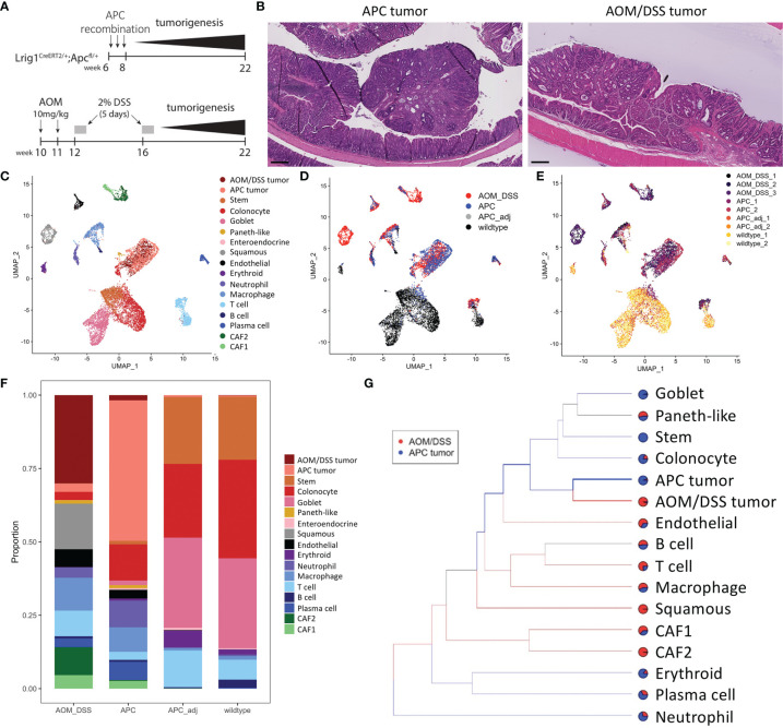
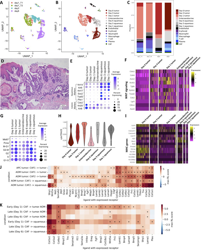
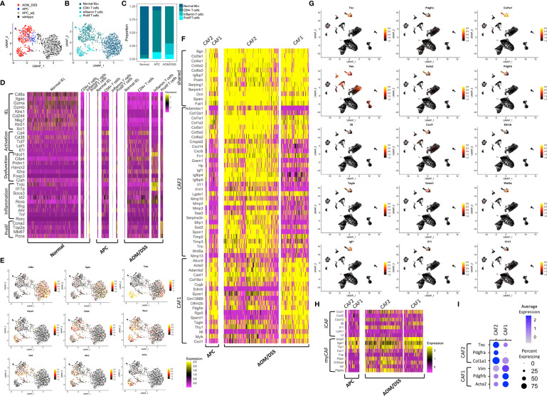
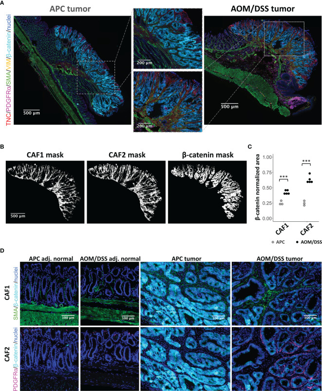
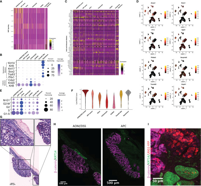
The tumor microenvironment plays a key role in the pathogenesis of colorectal tumors and contains various cell types including epithelial, immune, and mesenchymal cells. Characterization of the interactions between these cell types is necessary for revealing the complex nature of tumors. In this study, we used single-cell RNA-seq (scRNA-seq) to compare the tumor microenvironments between a mouse model of sporadic colorectal adenoma (Lrig1;Apc) and a mouse model of inflammation-driven colorectal cancer induced by azoxymethane and dextran sodium sulfate (AOM/DSS). While both models develop tumors in the distal colon, we found that the two tumor types have distinct microenvironments. AOM/DSS tumors have an increased abundance of two populations of cancer-associated fibroblasts (CAFs) compared with APC tumors, and we revealed their divergent spatial association with tumor cells using multiplex immunofluorescence (MxIF) imaging. We also identified a unique squamous cell population in AOM/DSS tumors, whose origins were distinct from anal squamous epithelial cells. These cells were in higher proportions upon administration of a chemotherapy regimen of 5-Fluorouracil/Irinotecan. We used computational inference algorithms to predict cell-cell communication mediated by ligand-receptor interactions and downstream pathway activation, and identified potential mechanistic connections between CAFs and tumor cells, as well as CAFs and squamous epithelial cells. This study provides important preclinical insight into the microenvironment of two distinct models of colorectal tumors and reveals unique roles for CAFs and squamous epithelial cells in the AOM/DSS model of inflammation-driven cancer.
肿瘤微环境在结直肠癌的发病机制中起关键作用,包含多种细胞类型,包括上皮细胞、免疫细胞和间充质细胞。表征这些细胞类型之间的相互作用对于揭示肿瘤的复杂本质至关重要。在本研究中,我们使用单细胞RNA测序(scRNA-seq)比较散发性结直肠腺瘤小鼠模型(Lrig1;Apc)和由氧化偶氮甲烷和葡聚糖硫酸钠诱导的炎症驱动型结直肠癌小鼠模型(AOM/DSS)之间的肿瘤微环境。虽然这两种模型均在远端结肠发生肿瘤,但我们发现这两种肿瘤类型具有不同的微环境。与APC肿瘤相比,AOM/DSS肿瘤中两种癌症相关成纤维细胞(CAF)群体的丰度增加,并且我们使用多重免疫荧光(MxIF)成像揭示了它们与肿瘤细胞的不同空间关联。我们还在AOM/DSS肿瘤中鉴定出一种独特的鳞状细胞群体,其起源与肛门鳞状上皮细胞不同。在给予5-氟尿嘧啶/伊立替康化疗方案后,这些细胞的比例更高。我们使用计算推理算法来预测由配体-受体相互作用和下游通路激活介导的细胞间通讯,并确定了CAF与肿瘤细胞以及CAF与鳞状上皮细胞之间潜在的机制联系。本研究为两种不同的结直肠癌模型的微环境提供了重要的临床前见解,并揭示了CAF和鳞状上皮细胞在炎症驱动型癌症的AOM/DSS模型中的独特作用。