Gambini Gloria, Carlà Matteo Mario, Giannuzzi Federico, Boselli Francesco, Grieco Giulia, Caporossi Tomaso, De Vico Umberto, Savastano Alfonso, Baldascino Antonio, Rizzo Clara, Kilian Raphael, Caporossi Aldo, Rizzo Stanislao
Ophthalmology Unit, Fondazione Policlinico Universitario A. Gemelli, IRCCS, 00168 Rome, Italy.
Ophthalmology Unit, Catholic University "Sacro Cuore", 00168 Rome, Italy.
Diagnostics (Basel). 2022 May 17;12(5):1250. doi: 10.3390/diagnostics12051250.
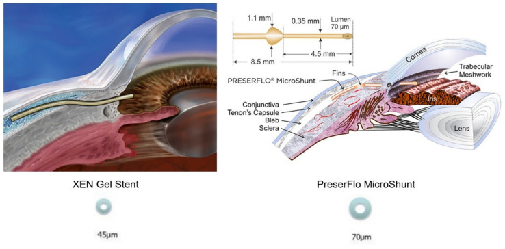
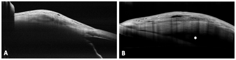
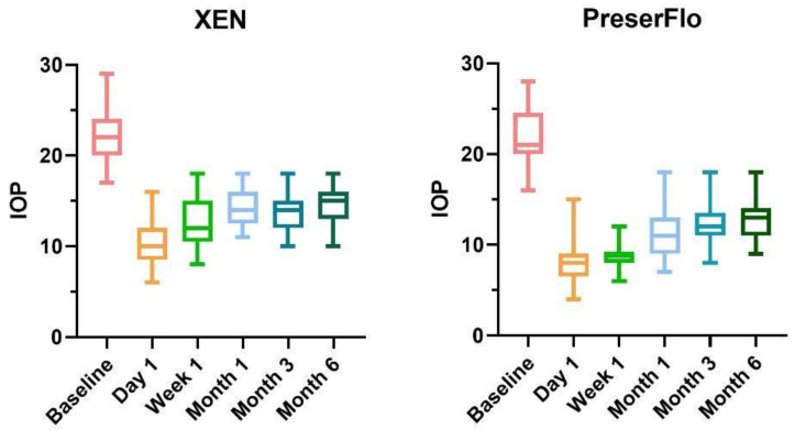
Background: The purpose of this study is to compare the morphology of six-month follow-up blebs created by a subconjunctival glaucoma surgical device (XEN45) to those created by a PreserFlo MicroShunt with a sub-Tenon insertion, utilizing AS-OCT. Methods: A retrospective study of 29 eyes who underwent XEN45 implantation and 29 eyes who underwent PreserFlo MicroShunt implantation. The patients were analyzed at 24 h, 1 week, 1 month, 3 months and 6 months. At each visit, the maturation and morphological alterations of the blebs were observed, as well as connections with the IOP. Results: In both groups, IOP showed significant reduction at all follow ups (p < 0.0001). In XEN group, the most common bleb morphology in the immediate postoperative was the subconjuntival separation type (42%) followed by the uniform type (34%), with a trend inversion at 6 month follow up (51% of uniform type). On the contrary, the most common morphology after PreserFlo was the multiple internal layer (55%), which showed a tendency to reduce over time and was substituted by the microcystic multiform, whose percentage increased over time (17% at day 1 vs. 44% at month 6). Uniform appearance was associated by the posterior episcleral fluid (PEF) lake presence. Both horizontal and vertical diameters significantly increased over time. Conclusion: XEN and PreserFlo implantation resulted in the production of diffuse blebs with different characteristics, which may influence IOP lowering capacity and bleb revisions necessity over time.
本研究的目的是利用AS-OCT比较结膜下青光眼手术装置(XEN45)与经Tenon囊下植入的PreserFlo微型分流器所形成的6个月随访期的滤泡形态。方法:对29例行XEN45植入术的眼和29例行PreserFlo微型分流器植入术的眼进行回顾性研究。在术后24小时、1周、1个月、3个月和6个月对患者进行分析。每次随访时,观察滤泡的成熟情况和形态改变,以及与眼压的关系。结果:两组在所有随访中眼压均显著降低(p<0.0001)。在XEN组,术后即刻最常见的滤泡形态是结膜下分离型(42%),其次是均匀型(34%),在6个月随访时趋势反转(均匀型占51%)。相反,PreserFlo术后最常见的形态是多层内层型(55%),其随时间有减少趋势,并被微囊多形型取代,微囊多形型的百分比随时间增加(第1天为17%,第6个月为44%)。均匀外观与巩膜后积液(PEF)湖的存在有关。水平和垂直直径均随时间显著增加。结论:XEN和PreserFlo植入导致产生具有不同特征的弥漫性滤泡,这可能会影响眼压降低能力以及随时间推移滤泡修复的必要性。