Li Jiaji, Zhou Ning, Sun Jiasong, Zhou Shun, Bai Zhidong, Lu Linpeng, Chen Qian, Zuo Chao
School of Electronic and Optical Engineering, Nanjing University of Science and Technology, No. 200 Xiaolingwei Street, Nanjing, Jiangsu Province, 210094, China.
Jiangsu Key Laboratory of Spectral Imaging & Intelligent Sense, Nanjing University of Science and Technology, Nanjing, Jiangsu Province, 210094, China.
Light Sci Appl. 2022 Jun 2;11(1):154. doi: 10.1038/s41377-022-00815-7.
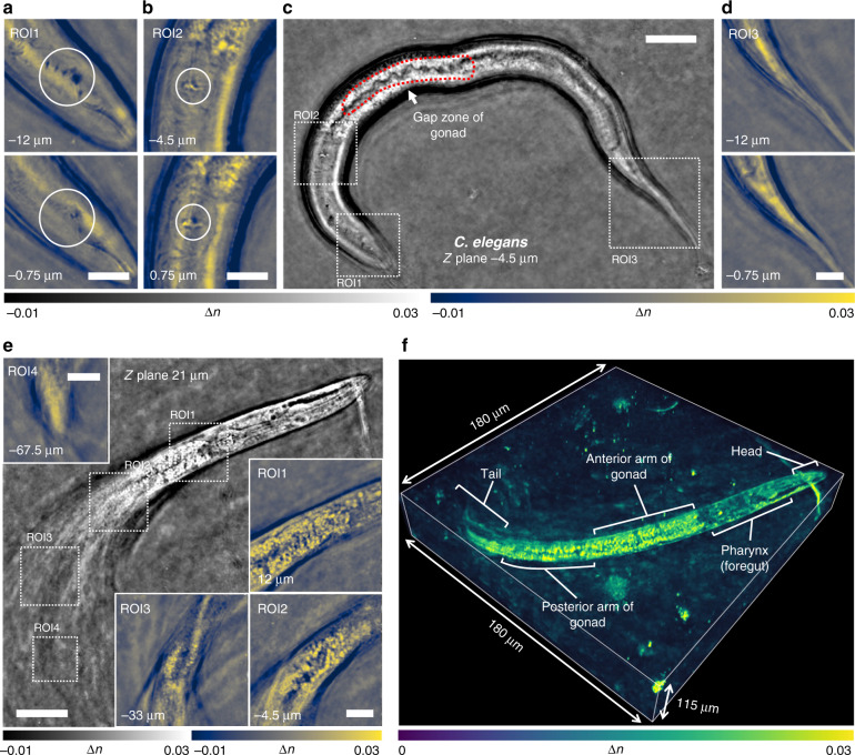
We present a new label-free three-dimensional (3D) microscopy technique, termed transport of intensity diffraction tomography with non-interferometric synthetic aperture (TIDT-NSA). Without resorting to interferometric detection, TIDT-NSA retrieves the 3D refractive index (RI) distribution of biological specimens from 3D intensity-only measurements at various illumination angles, allowing incoherent-diffraction-limited quantitative 3D phase-contrast imaging. The unique combination of z-scanning the sample with illumination angle diversity in TIDT-NSA provides strong defocus phase contrast and better optical sectioning capabilities suitable for high-resolution tomography of thick biological samples. Based on an off-the-shelf bright-field microscope with a programmable light-emitting-diode (LED) illumination source, TIDT-NSA achieves an imaging resolution of 206 nm laterally and 520 nm axially with a high-NA oil immersion objective. We validate the 3D RI tomographic imaging performance on various unlabeled fixed and live samples, including human breast cancer cell lines MCF-7, human hepatocyte carcinoma cell lines HepG2, mouse macrophage cell lines RAW 264.7, Caenorhabditis elegans (C. elegans), and live Henrietta Lacks (HeLa) cells. These results establish TIDT-NSA as a new non-interferometric approach to optical diffraction tomography and 3D label-free microscopy, permitting quantitative characterization of cell morphology and time-dependent subcellular changes for widespread biological and medical applications.
我们提出了一种新的无标记三维(3D)显微镜技术,称为非干涉合成孔径强度传输衍射层析成像(TIDT-NSA)。TIDT-NSA无需干涉检测,而是通过在不同照明角度下进行仅强度的3D测量来获取生物样本的3D折射率(RI)分布,从而实现非相干衍射极限的定量3D相衬成像。TIDT-NSA中对样品进行z扫描并结合照明角度多样性,这种独特的组合提供了强大的离焦相衬和更好的光学切片能力,适用于厚生物样本的高分辨率层析成像。基于一台配备可编程发光二极管(LED)照明源的现成明场显微镜,TIDT-NSA使用高数值孔径油浸物镜实现了横向206 nm和轴向520 nm的成像分辨率。我们在各种未标记的固定和活样本上验证了3D RI层析成像性能,包括人乳腺癌细胞系MCF-7、人肝癌细胞系HepG2、小鼠巨噬细胞系RAW 264.7、秀丽隐杆线虫(C. elegans)以及活的海拉(HeLa)细胞。这些结果确立了TIDT-NSA作为一种新的非干涉光学衍射层析成像和3D无标记显微镜方法,能够对细胞形态和随时间变化的亚细胞变化进行定量表征,适用于广泛的生物学和医学应用。