Dong Dashan, Huang Xiaoshuai, Li Liuju, Mao Heng, Mo Yanquan, Zhang Guangyi, Zhang Zhe, Shen Jiayu, Liu Wei, Wu Zeming, Liu Guanghui, Liu Yanmei, Yang Hong, Gong Qihuang, Shi Kebin, Chen Liangyi
1State Key Laboratory for Mesoscopic Physics and Frontiers Science Center for Nano-optoelectronics, School of Physics, Peking University, Beijing, 100871 China.
2Collaborative Innovation Center of Extreme Optics, Shanxi University, Taiyuan, Shanxi 030006 China.
Light Sci Appl. 2020 Jan 28;9:11. doi: 10.1038/s41377-020-0249-4. eCollection 2020.
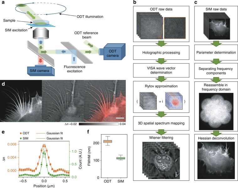
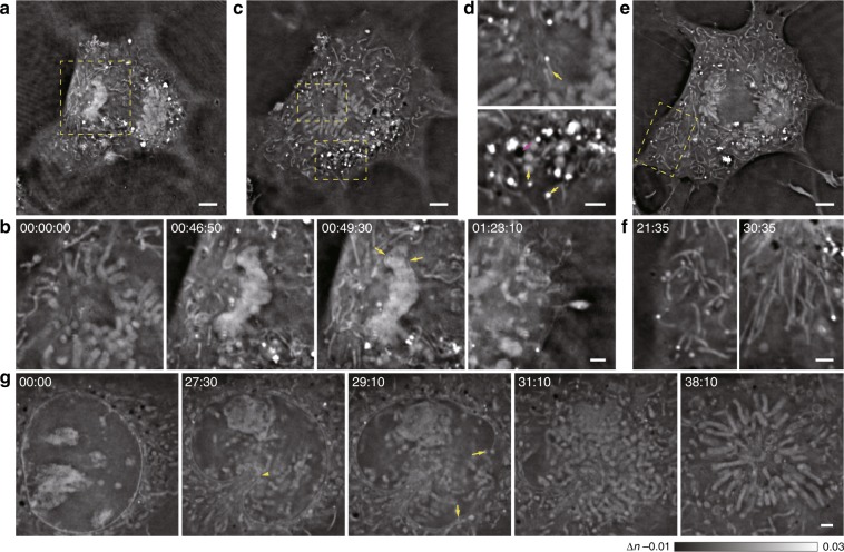
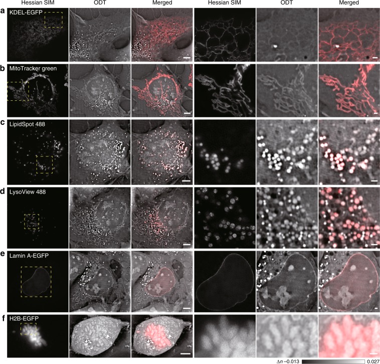
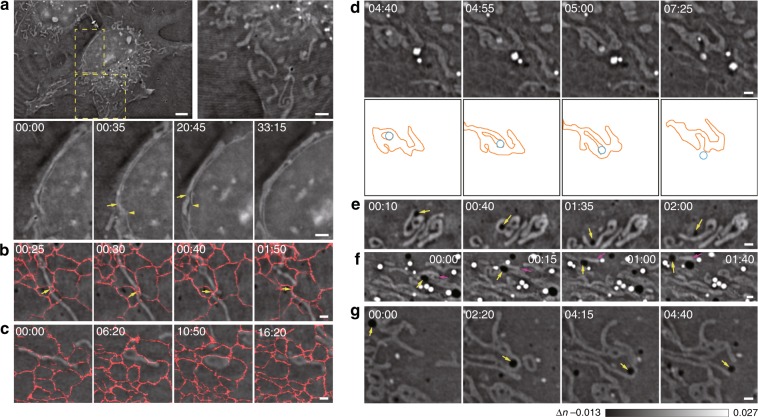
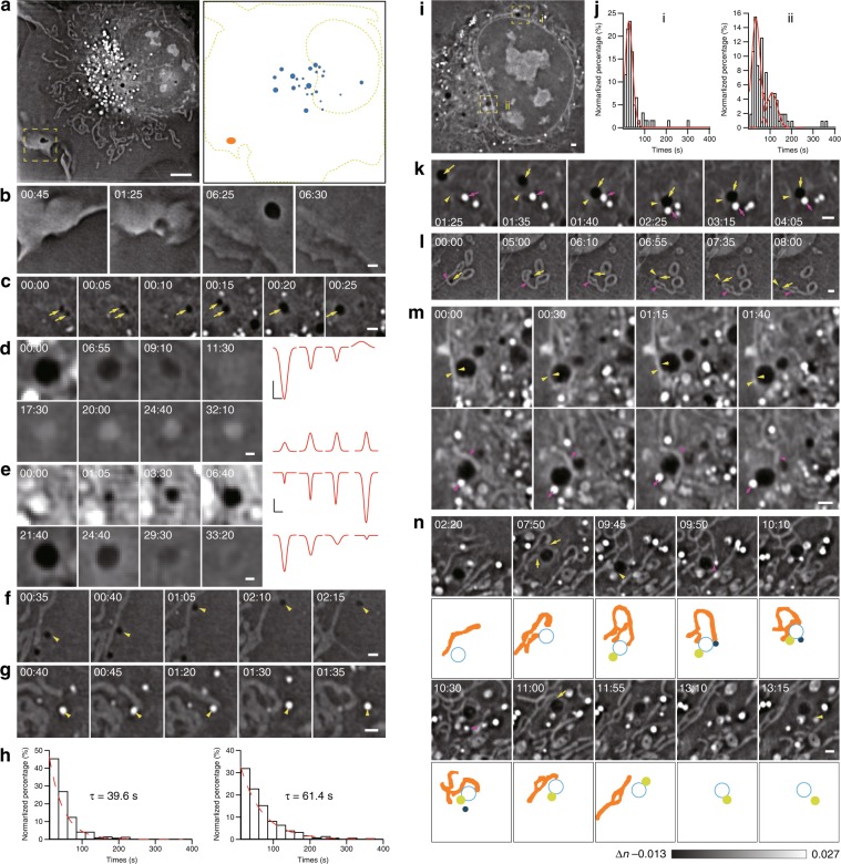
The emergence of super-resolution (SR) fluorescence microscopy has rejuvenated the search for new cellular sub-structures. However, SR fluorescence microscopy achieves high contrast at the expense of a holistic view of the interacting partners and surrounding environment. Thus, we developed SR fluorescence-assisted diffraction computational tomography (SR-FACT), which combines label-free three-dimensional optical diffraction tomography (ODT) with two-dimensional fluorescence Hessian structured illumination microscopy. The ODT module is capable of resolving the mitochondria, lipid droplets, the nuclear membrane, chromosomes, the tubular endoplasmic reticulum, and lysosomes. Using dual-mode correlated live-cell imaging for a prolonged period of time, we observed novel subcellular structures named dark-vacuole bodies, the majority of which originate from densely populated perinuclear regions, and intensively interact with organelles such as the mitochondria and the nuclear membrane before ultimately collapsing into the plasma membrane. This work demonstrates the unique capabilities of SR-FACT, which suggests its wide applicability in cell biology in general.
超分辨率(SR)荧光显微镜的出现为寻找新的细胞亚结构注入了新活力。然而,SR荧光显微镜是以牺牲对相互作用伙伴及周围环境的整体观察为代价来实现高对比度的。因此,我们开发了SR荧光辅助衍射计算断层扫描(SR-FACT),它将无标记三维光学衍射断层扫描(ODT)与二维荧光海森结构照明显微镜相结合。ODT模块能够分辨线粒体、脂滴、核膜、染色体、管状内质网和溶酶体。通过长时间的双模式相关活细胞成像,我们观察到了名为暗空泡体的新型亚细胞结构,其中大多数起源于密集的核周区域,并在最终塌陷到质膜之前与线粒体和核膜等细胞器强烈相互作用。这项工作展示了SR-FACT的独特能力,这表明它在细胞生物学中具有广泛的适用性。