• 文献检索
  • 文档翻译
  • 深度研究
  • 学术资讯
  • Suppr Zotero 插件Zotero 插件
  • 邀请有礼
  • 套餐&价格
  • 历史记录
应用&插件
Suppr Zotero 插件Zotero 插件浏览器插件Mac 客户端Windows 客户端微信小程序
定价
高级版会员购买积分包购买API积分包
服务
文献检索文档翻译深度研究API 文档MCP 服务
关于我们
关于 Suppr公司介绍联系我们用户协议隐私条款
关注我们

Suppr 超能文献

核心技术专利:CN118964589B侵权必究
粤ICP备2023148730 号-1Suppr @ 2026

文献检索

告别复杂PubMed语法,用中文像聊天一样搜索,搜遍4000万医学文献。AI智能推荐,让科研检索更轻松。

立即免费搜索

文件翻译

保留排版,准确专业,支持PDF/Word/PPT等文件格式,支持 12+语言互译。

免费翻译文档

深度研究

AI帮你快速写综述,25分钟生成高质量综述,智能提取关键信息,辅助科研写作。

立即免费体验

原发性侧索硬化症中胼胝体区 III 的局灶性改变:MRI 平面测量和纹理分析。

Focal alterations of the callosal area III in primary lateral sclerosis: An MRI planimetry and texture analysis.

机构信息

Department of Neurology, University of Ulm, Germany.

Institute of Epidemiology and Medical Biometry, University of Ulm, Germany.

出版信息

Neuroimage Clin. 2020;26:102223. doi: 10.1016/j.nicl.2020.102223. Epub 2020 Feb 21.

DOI:10.1016/j.nicl.2020.102223
PMID:32114375
原文链接:https://pmc.ncbi.nlm.nih.gov/articles/PMC7049663/
Abstract

BACKGROUND

The regional distribution of cerebral morphological alterations in primary lateral sclerosis (PLS) is considered to include the area III of the corpus callosum (CC).

OBJECTIVE

The study was designed to investigate regional white matter (WM) alterations in the callosal area III by T1 weighted magnetic resonance imaging (T1w-MRI) data in PLS patients compared with healthy controls, in order to identify atrophy and texture changes in vivo.

METHODS

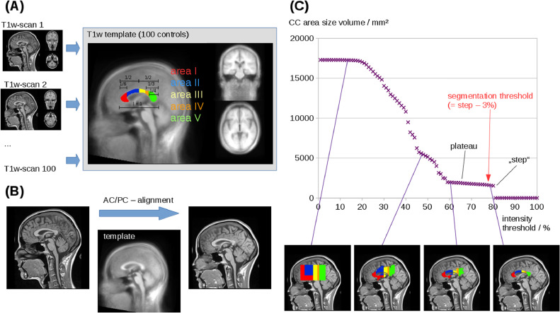
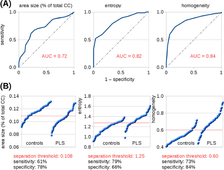
T1w-MRI-based white matter mapping was used to perform an operator-independent CC-segmentation for the different areas of the CC in 67 PLS patients vs 82 matched healthy controls and vs 85 ALS patients. The segmentation was followed by texture analysis of the separated CC areas for the PLS patients vs controls and vs ALS patients.

RESULTS

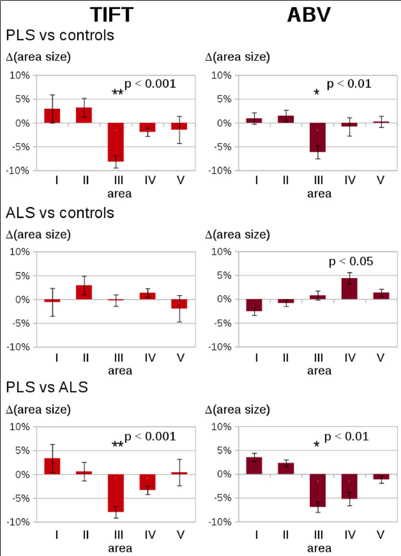
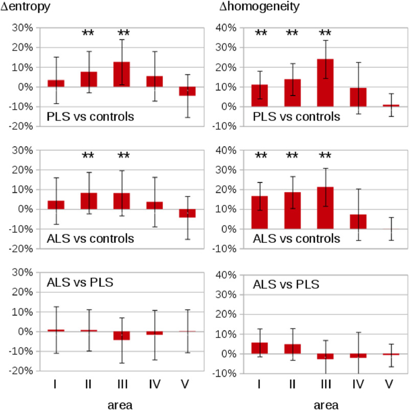
PLS was associated with significant atrophy in the area III of the CC (but not in the other callosal segments), while the alterations in the ALS patients were much more variable and were not significant at the group level. Furthermore, significant regional alterations of the texture parameters entropy and homogeneity in this area were shown in PLS patients and in ALS patients.

CONCLUSIONS

This T1w-MRI study demonstrated focused regional CC atrophy and texture alterations limited to the callosal area III (which comprises fibers projecting into the primary motor cortices) in PLS, in comparison to a higher variability in CC size in ALS.

摘要

背景

原发性侧索硬化症(PLS)的脑形态改变的区域分布被认为包括胼胝体(CC)的区域 III。

目的

本研究旨在通过与健康对照组相比,在 PLS 患者中调查 CC 区域 III 的白质(WM)改变,以在体内识别萎缩和纹理变化。

方法

使用 T1 加权磁共振成像(T1w-MRI)数据对 67 名 PLS 患者与 82 名匹配的健康对照组和 85 名 ALS 患者进行 T1w-MRI 基于白质的映射,以执行不同的 CC 区域的独立操作者的 CC 分割。对 PLS 患者与对照组和 ALS 患者进行分离的 CC 区域的纹理分析。

结果

PLS 与 CC 区域 III 的明显萎缩相关(但与其他胼胝体段无关),而 ALS 患者的改变则更加多变,且在组水平上无显著差异。此外,在该区域显示出纹理参数熵和同质性的明显区域改变,在 PLS 患者和 ALS 患者中。

结论

这项 T1w-MRI 研究表明,与 ALS 相比,PLS 中胼胝体的特定区域萎缩和纹理改变仅限于 CC 区域 III(包括投射到初级运动皮质的纤维),而 CC 大小的变异性更高。

https://cdn.ncbi.nlm.nih.gov/pmc/blobs/9b83/7049663/887918abc596/gr5.jpg
https://cdn.ncbi.nlm.nih.gov/pmc/blobs/9b83/7049663/89bcf6a60bf0/gr1.jpg
https://cdn.ncbi.nlm.nih.gov/pmc/blobs/9b83/7049663/05c78af23911/gr2.jpg
https://cdn.ncbi.nlm.nih.gov/pmc/blobs/9b83/7049663/ced2e35d6e01/gr3.jpg
https://cdn.ncbi.nlm.nih.gov/pmc/blobs/9b83/7049663/be094cf9ae0c/gr4.jpg
https://cdn.ncbi.nlm.nih.gov/pmc/blobs/9b83/7049663/887918abc596/gr5.jpg

相似文献

1
Focal alterations of the callosal area III in primary lateral sclerosis: An MRI planimetry and texture analysis.原发性侧索硬化症中胼胝体区 III 的局灶性改变:MRI 平面测量和纹理分析。
Neuroimage Clin. 2020;26:102223. doi: 10.1016/j.nicl.2020.102223. Epub 2020 Feb 21.
2
Segmental alterations of the corpus callosum in motor neuron disease: A DTI and texture analysis in 575 patients.运动神经元病胼胝体节段性改变:575 例患者的 DTI 和纹理分析。
Neuroimage Clin. 2022;35:103061. doi: 10.1016/j.nicl.2022.103061. Epub 2022 May 28.
3
Brainstem pathology in amyotrophic lateral sclerosis and primary lateral sclerosis: A longitudinal neuroimaging study.肌萎缩侧索硬化症和原发性侧索硬化症的脑干病理学:一项纵向神经影像学研究。
Neuroimage Clin. 2019;24:102054. doi: 10.1016/j.nicl.2019.102054. Epub 2019 Oct 24.
4
Identical patterns of cortico-efferent tract involvement in primary lateral sclerosis and amyotrophic lateral sclerosis: A tract of interest-based MRI study.原发性侧索硬化症和肌萎缩侧索硬化症皮质传出束受累的相同模式:基于感兴趣区的磁共振成像研究。
Neuroimage Clin. 2018 Mar 15;18:762-769. doi: 10.1016/j.nicl.2018.03.018. eCollection 2018.
5
Segmental Alterations of the Corpus Callosum in Progressive Supranuclear Palsy: A Multiparametric Magnetic Resonance Imaging Study.进行性核上性麻痹中胼胝体的节段性改变:一项多参数磁共振成像研究
Front Aging Neurosci. 2021 Nov 19;13:720634. doi: 10.3389/fnagi.2021.720634. eCollection 2021.
6
Intrahemispheric and interhemispheric structural network abnormalities in PLS and ALS.进行性核上性麻痹和肌萎缩侧索硬化症患者的半球内和半球间结构网络异常。
Hum Brain Mapp. 2014 Apr;35(4):1710-22. doi: 10.1002/hbm.22286. Epub 2013 Apr 30.
7
Cortico-efferent tract involvement in primary lateral sclerosis and amyotrophic lateral sclerosis: A two-centre tract of interest-based DTI analysis.皮质传出束在原发性侧索硬化症和肌萎缩侧索硬化症中的受累:基于两中心感兴趣区的 DTI 分析。
Neuroimage Clin. 2018;20:1062-1069. doi: 10.1016/j.nicl.2018.10.005. Epub 2018 Oct 9.
8
Cross-sectional and longitudinal assessment of the upper cervical spinal cord in motor neuron disease.运动神经元病中颈上脊髓的横断和纵向评估。
Neuroimage Clin. 2019;24:101984. doi: 10.1016/j.nicl.2019.101984. Epub 2019 Aug 16.
9
Pathology of callosal damage in ALS: An , 7 T diffusion tensor MRI study.肌萎缩侧索硬化症胼胝体损伤的病理学:一项7T扩散张量磁共振成像研究
Neuroimage Clin. 2017 Apr 30;15:200-208. doi: 10.1016/j.nicl.2017.04.024. eCollection 2017.
10
White matter alterations differ in primary lateral sclerosis and amyotrophic lateral sclerosis.白质改变在原发性侧索硬化症和肌萎缩性侧索硬化症中有所不同。
Brain. 2011 Sep;134(Pt 9):2642-55. doi: 10.1093/brain/awr178. Epub 2011 Jul 28.

引用本文的文献

1
Mapping motor and extra-motor gray and white matter changes in ALS: a comprehensive review of MRI insights.肌萎缩侧索硬化症中运动和运动外灰质及白质变化的图谱绘制:MRI研究见解的全面综述
Neuroradiology. 2025 May 2. doi: 10.1007/s00234-025-03629-7.
2
Neuroimaging findings in preclinical amyotrophic lateral sclerosis models-How well do they mimic the clinical phenotype? A systematic review.临床前肌萎缩侧索硬化模型的神经影像学研究结果——它们对临床表型的模拟效果如何?一项系统综述。
Front Vet Sci. 2023 May 2;10:1135282. doi: 10.3389/fvets.2023.1135282. eCollection 2023.
3
Structural magnetic resonance imaging findings and histopathological correlations in motor neuron diseases-A systematic review and meta-analysis.

本文引用的文献

1
Brainstem pathology in amyotrophic lateral sclerosis and primary lateral sclerosis: A longitudinal neuroimaging study.肌萎缩侧索硬化症和原发性侧索硬化症的脑干病理学:一项纵向神经影像学研究。
Neuroimage Clin. 2019;24:102054. doi: 10.1016/j.nicl.2019.102054. Epub 2019 Oct 24.
2
Corticospinal tract degeneration in ALS unmasked in T1-weighted images using texture analysis.使用纹理分析在 T1 加权图像中揭示 ALS 中的皮质脊髓束退变。
Hum Brain Mapp. 2019 Mar;40(4):1174-1183. doi: 10.1002/hbm.24437. Epub 2018 Oct 27.
3
Cortico-efferent tract involvement in primary lateral sclerosis and amyotrophic lateral sclerosis: A two-centre tract of interest-based DTI analysis.
运动神经元疾病的结构磁共振成像结果与组织病理学相关性——一项系统评价和荟萃分析
Front Neurol. 2022 Aug 30;13:947347. doi: 10.3389/fneur.2022.947347. eCollection 2022.
4
Multiparametric Microstructural MRI and Machine Learning Classification Yields High Diagnostic Accuracy in Amyotrophic Lateral Sclerosis: Proof of Concept.多参数微观结构磁共振成像与机器学习分类在肌萎缩侧索硬化症中具有高诊断准确性:概念验证
Front Neurol. 2021 Nov 17;12:745475. doi: 10.3389/fneur.2021.745475. eCollection 2021.
5
Segmental Alterations of the Corpus Callosum in Progressive Supranuclear Palsy: A Multiparametric Magnetic Resonance Imaging Study.进行性核上性麻痹中胼胝体的节段性改变:一项多参数磁共振成像研究
Front Aging Neurosci. 2021 Nov 19;13:720634. doi: 10.3389/fnagi.2021.720634. eCollection 2021.
6
Feature selection from magnetic resonance imaging data in ALS: a systematic review.肌萎缩侧索硬化症中磁共振成像数据的特征选择:一项系统综述
Ther Adv Chronic Dis. 2021 Oct 13;12:20406223211051002. doi: 10.1177/20406223211051002. eCollection 2021.
7
The ipsilateral silent period: an early diagnostic marker of callosal disconnection in ALS.同侧静息期:肌萎缩侧索硬化症胼胝体离断的早期诊断标志物。
Ther Adv Chronic Dis. 2021 Sep 13;12:20406223211044072. doi: 10.1177/20406223211044072. eCollection 2021.
8
Segmental involvement of the corpus callosum in associated ALS: a tract of interest-based DTI study.肌萎缩侧索硬化症相关病例中胼胝体的节段性受累:一项基于感兴趣区的扩散张量成像研究
Ther Adv Chronic Dis. 2021 Mar 21;12:20406223211002969. doi: 10.1177/20406223211002969. eCollection 2021.
皮质传出束在原发性侧索硬化症和肌萎缩侧索硬化症中的受累:基于两中心感兴趣区的 DTI 分析。
Neuroimage Clin. 2018;20:1062-1069. doi: 10.1016/j.nicl.2018.10.005. Epub 2018 Oct 9.
4
Identical patterns of cortico-efferent tract involvement in primary lateral sclerosis and amyotrophic lateral sclerosis: A tract of interest-based MRI study.原发性侧索硬化症和肌萎缩侧索硬化症皮质传出束受累的相同模式:基于感兴趣区的磁共振成像研究。
Neuroimage Clin. 2018 Mar 15;18:762-769. doi: 10.1016/j.nicl.2018.03.018. eCollection 2018.
5
Multimodal structural MRI in the diagnosis of motor neuron diseases.多模态结构磁共振成像在运动神经元疾病诊断中的应用
Neuroimage Clin. 2017 Aug 2;16:240-247. doi: 10.1016/j.nicl.2017.08.002. eCollection 2017.
6
Virtual brain biopsies in amyotrophic lateral sclerosis: Diagnostic classification based on in vivo pathological patterns.肌萎缩侧索硬化症的虚拟脑活检:基于体内病理模式的诊断分类
Neuroimage Clin. 2017 Jun 9;15:653-658. doi: 10.1016/j.nicl.2017.06.010. eCollection 2017.
7
The concept and diagnostic criteria of primary lateral sclerosis.原发性侧索硬化症的概念及诊断标准。
Acta Neurol Scand. 2017 Sep;136(3):204-211. doi: 10.1111/ane.12713. Epub 2016 Nov 15.
8
Cerebral Degeneration in Amyotrophic Lateral Sclerosis Revealed by 3-Dimensional Texture Analysis.三维纹理分析揭示肌萎缩侧索硬化症中的脑变性
Front Neurosci. 2016 Mar 30;10:120. doi: 10.3389/fnins.2016.00120. eCollection 2016.
9
A large-scale multicentre cerebral diffusion tensor imaging study in amyotrophic lateral sclerosis.一项大型多中心肌萎缩侧索硬化症的脑弥散张量成像研究。
J Neurol Neurosurg Psychiatry. 2016 Jun;87(6):570-9. doi: 10.1136/jnnp-2015-311952. Epub 2016 Jan 8.
10
A revision of the El Escorial criteria - 2015.《埃尔埃斯科里亚尔标准(2015年修订版)》
Amyotroph Lateral Scler Frontotemporal Degener. 2015;16(5-6):291-2. doi: 10.3109/21678421.2015.1049183. Epub 2015 Jun 29.