• 文献检索
  • 文档翻译
  • 深度研究
  • 学术资讯
  • Suppr Zotero 插件Zotero 插件
  • 邀请有礼
  • 套餐&价格
  • 历史记录
应用&插件
Suppr Zotero 插件Zotero 插件浏览器插件Mac 客户端Windows 客户端微信小程序
定价
高级版会员购买积分包购买API积分包
服务
文献检索文档翻译深度研究API 文档MCP 服务
关于我们
关于 Suppr公司介绍联系我们用户协议隐私条款
关注我们

Suppr 超能文献

核心技术专利:CN118964589B侵权必究
粤ICP备2023148730 号-1Suppr @ 2026

文献检索

告别复杂PubMed语法,用中文像聊天一样搜索,搜遍4000万医学文献。AI智能推荐,让科研检索更轻松。

立即免费搜索

文件翻译

保留排版,准确专业,支持PDF/Word/PPT等文件格式,支持 12+语言互译。

免费翻译文档

深度研究

AI帮你快速写综述,25分钟生成高质量综述,智能提取关键信息,辅助科研写作。

立即免费体验

一个整个人体左肺的多尺度 X 射线相衬层析数据集。

A multiscale X-ray phase-contrast tomography dataset of a whole human left lung.

机构信息

Department of Mechanical Engineering, University College London, London, UK.

Antwerp Surgical Training, Anatomy and Research Centre (ASTARC), University of Antwerp, Wilrijk, Belgium.

出版信息

Sci Data. 2022 Jun 2;9(1):264. doi: 10.1038/s41597-022-01353-y.

DOI:10.1038/s41597-022-01353-y
PMID:35654864
原文链接:https://pmc.ncbi.nlm.nih.gov/articles/PMC9163096/
Abstract

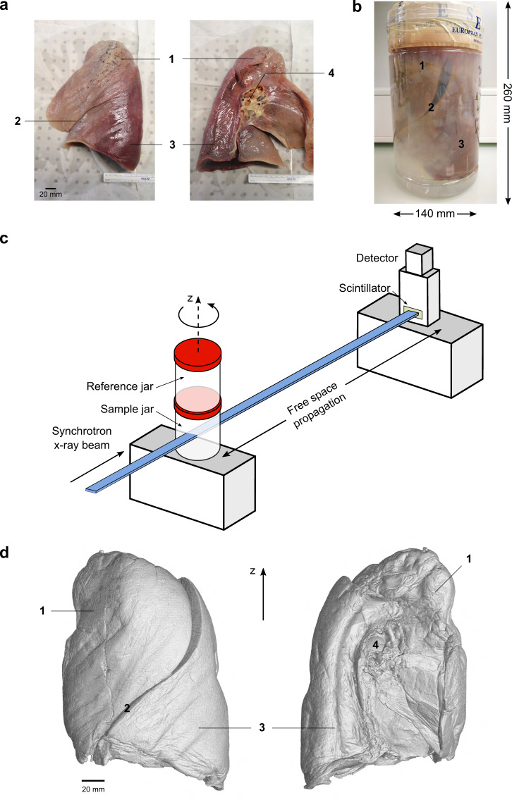
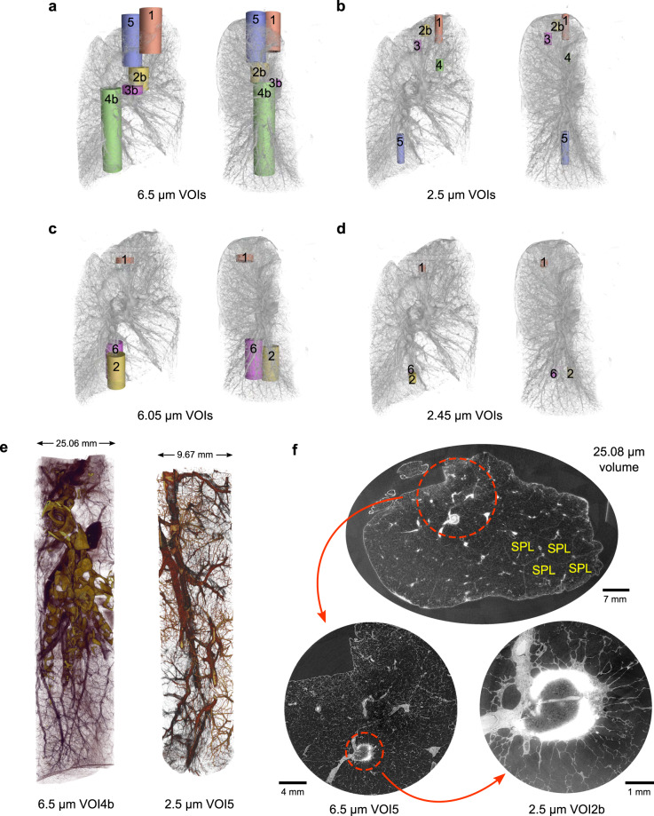
Technological advancements in X-ray imaging using bright and coherent synchrotron sources now allows the decoupling of sample size and resolution while maintaining high sensitivity to the microstructures of soft, partially dehydrated tissues. The continuous developments in multiscale X-ray imaging resulted in hierarchical phase-contrast tomography, a comprehensive approach to address the challenge of organ-scale (up to tens of centimeters) soft tissue imaging with resolution and sensitivity down to the cellular level. Using this technique, we imaged ex vivo an entire human left lung at an isotropic voxel size of 25.08 μm along with local zooms down to 6.05-6.5 μm and 2.45-2.5 μm in voxel size. The high tissue contrast offered by the fourth-generation synchrotron source at the European Synchrotron Radiation Facility reveals the complex multiscale anatomical constitution of the human lung from the macroscopic (centimeter) down to the microscopic (micrometer) scale. The dataset provides comprehensive organ-scale 3D information of the secondary pulmonary lobules and delineates the microstructure of lung nodules with unprecedented detail.

摘要

利用明亮且相干的同步辐射源的 X 射线成像技术的进步,现在可以在保持对软、部分脱水组织微观结构的高灵敏度的同时,解耦样品尺寸和分辨率。多尺度 X 射线成像的不断发展导致了分级相衬断层摄影术的出现,这是一种全面的方法,可以解决器官尺度(可达数十厘米)软组织成像的挑战,分辨率和灵敏度可达到细胞水平。使用这种技术,我们对整个人体左肺进行了离体成像,体素大小为 25.08μm,局部放大倍数分别为 6.05-6.5μm 和 2.45-2.5μm。欧洲同步辐射装置第四代同步辐射源提供的高组织对比度揭示了人体肺从宏观(厘米)到微观(微米)尺度的复杂多尺度解剖结构。该数据集提供了次级肺小叶的全面器官尺度 3D 信息,并以前所未有的细节描绘了肺结节的微观结构。

https://cdn.ncbi.nlm.nih.gov/pmc/blobs/6783/9163096/f165a89ee4ef/41597_2022_1353_Fig3_HTML.jpg
https://cdn.ncbi.nlm.nih.gov/pmc/blobs/6783/9163096/61e282cba549/41597_2022_1353_Fig1_HTML.jpg
https://cdn.ncbi.nlm.nih.gov/pmc/blobs/6783/9163096/ddb9dab5bc0b/41597_2022_1353_Fig2_HTML.jpg
https://cdn.ncbi.nlm.nih.gov/pmc/blobs/6783/9163096/f165a89ee4ef/41597_2022_1353_Fig3_HTML.jpg
https://cdn.ncbi.nlm.nih.gov/pmc/blobs/6783/9163096/61e282cba549/41597_2022_1353_Fig1_HTML.jpg
https://cdn.ncbi.nlm.nih.gov/pmc/blobs/6783/9163096/ddb9dab5bc0b/41597_2022_1353_Fig2_HTML.jpg
https://cdn.ncbi.nlm.nih.gov/pmc/blobs/6783/9163096/f165a89ee4ef/41597_2022_1353_Fig3_HTML.jpg

相似文献

1
A multiscale X-ray phase-contrast tomography dataset of a whole human left lung.一个整个人体左肺的多尺度 X 射线相衬层析数据集。
Sci Data. 2022 Jun 2;9(1):264. doi: 10.1038/s41597-022-01353-y.
2
Multidimensional Analysis of the Adult Human Heart in Health and Disease Using Hierarchical Phase-Contrast Tomography.采用分层相位对比层析成像技术对健康和疾病状态下成人心脏的多维分析。
Radiology. 2024 Jul;312(1):e232731. doi: 10.1148/radiol.232731.
3
Human lung virtual histology by multi-scale x-ray phase-contrast computed tomography.多尺度 X 射线相衬计算机断层扫描的人类肺部虚拟组织学。
Phys Med Biol. 2023 May 30;68(11). doi: 10.1088/1361-6560/acd48d.
4
Preparation of large biological samples for high-resolution, hierarchical, synchrotron phase-contrast tomography with multimodal imaging compatibility.为具有多模态成像兼容性的高分辨率、层次化同步相衬断层摄影术准备大型生物样本。
Nat Protoc. 2023 May;18(5):1441-1461. doi: 10.1038/s41596-023-00804-z. Epub 2023 Mar 1.
5
Imaging performance of phase-contrast breast computed tomography with synchrotron radiation and a CdTe photon-counting detector.相衬乳腺计算机断层成像的同步辐射成像性能与碲锌镉光子计数探测器
Phys Med. 2016 May;32(5):681-90. doi: 10.1016/j.ejmp.2016.04.011. Epub 2016 May 6.
6
Synchrotron X-ray imaging of soft biological tissues - principles, applications and future prospects.同步辐射 X 射线成像技术在软生物组织中的应用——原理、应用和未来展望。
J Cell Sci. 2024 Oct 15;137(20). doi: 10.1242/jcs.261953. Epub 2024 Oct 23.
7
Multiscale three-dimensional imaging of intact human organs down to the cellular scale using hierarchical phase-contrast tomography.使用分层相衬断层扫描技术对完整人体器官进行直至细胞尺度的多尺度三维成像。
bioRxiv. 2021 Feb 3:2021.02.03.429481. doi: 10.1101/2021.02.03.429481.
8
Visualization Ability of Phase-Contrast Synchrotron-Based X-Ray Imaging Using an X-Ray Interferometer in Soft Tissue Tumors.基于相衬同步辐射 X 射线干涉仪的软组织肿瘤 X 射线成像的可视化能力。
Technol Cancer Res Treat. 2021 Jan-Dec;20:15330338211010121. doi: 10.1177/15330338211010121.
9
Potential of propagation-based synchrotron X-ray phase-contrast computed tomography for cardiac tissue engineering.基于传播的同步加速器X射线相衬计算机断层扫描在心脏组织工程中的潜力。
J Synchrotron Radiat. 2017 Jul 1;24(Pt 4):842-853. doi: 10.1107/S1600577517006208. Epub 2017 May 18.
10
Three-dimensional construction of micrometer level in rat stomach by synchrotron radiation.利用同步辐射对大鼠胃进行微米级三维构建。
Biomed Eng Online. 2021 Mar 20;20(1):28. doi: 10.1186/s12938-021-00866-8.

引用本文的文献

1
Measurements of source emittance and beam coherence properties of the upgraded Advanced Photon Source.升级后的先进光子源的源发射度和束流相干特性测量。
J Synchrotron Radiat. 2025 Sep 1;32(Pt 5):1152-1161. doi: 10.1107/S160057752500579X. Epub 2025 Aug 15.
2
Tube2FEM: a general-purpose highly automated pipeline for flow-related processes in (embedded) tubular objects.Tube2FEM:用于(嵌入式)管状物体中与流动相关过程的通用高度自动化管道。
R Soc Open Sci. 2025 Aug 13;12(8):242025. doi: 10.1098/rsos.242025. eCollection 2025 Aug.
3
3D imaging of the human temporal bone by X-ray phase-contrast tomography.

本文引用的文献

1
Opportunities for biomineralization research using multiscale computed X-ray tomography as exemplified by bone imaging.以骨成像为例,利用多尺度计算机X射线断层扫描进行生物矿化研究的机遇。
J Struct Biol. 2022 Mar;214(1):107822. doi: 10.1016/j.jsb.2021.107822. Epub 2021 Dec 10.
2
Imaging intact human organs with local resolution of cellular structures using hierarchical phase-contrast tomography.利用分层相衬断层摄影术,以局部分辨率对完整的人体器官进行成像。
Nat Methods. 2021 Dec;18(12):1532-1541. doi: 10.1038/s41592-021-01317-x. Epub 2021 Nov 4.
3
Hierarchical imaging and computational analysis of three-dimensional vascular network architecture in the entire postnatal and adult mouse brain.
通过X射线相衬断层扫描对人类颞骨进行三维成像。
Npj Imaging. 2025 May 20;3(1):21. doi: 10.1038/s44303-025-00086-y.
4
Deep learning for 3D vascular segmentation in hierarchical phase contrast tomography: a case study on kidney.深度学习在分层相衬断层摄影中的 3D 血管分割:以肾脏为例的研究
Sci Rep. 2024 Nov 8;14(1):27258. doi: 10.1038/s41598-024-77582-5.
5
Computer vision applications for the detection or analysis of tuberculosis using digitised human lung tissue images - a systematic review.基于数字化人肺组织图像的计算机视觉技术在结核病检测或分析中的应用:系统综述。
BMC Med Imaging. 2024 Nov 5;24(1):298. doi: 10.1186/s12880-024-01443-w.
6
Real-world federated learning in radiology: hurdles to overcome and benefits to gain.放射学中的真实世界联邦学习:需克服的障碍与可获得的益处
J Am Med Inform Assoc. 2025 Jan 1;32(1):193-205. doi: 10.1093/jamia/ocae259.
7
Synchrotron X-ray imaging of soft biological tissues - principles, applications and future prospects.同步辐射 X 射线成像技术在软生物组织中的应用——原理、应用和未来展望。
J Cell Sci. 2024 Oct 15;137(20). doi: 10.1242/jcs.261953. Epub 2024 Oct 23.
8
3D digital light process bioprinting: Cutting-edge platforms for resolution of organ fabrication.3D数字光处理生物打印:用于解决器官制造问题的前沿平台。
Mater Today Bio. 2024 Oct 2;29:101284. doi: 10.1016/j.mtbio.2024.101284. eCollection 2024 Dec.
9
Deep Learning for 3D Vascular Segmentation in Phase Contrast Tomography.用于相位对比断层扫描中三维血管分割的深度学习
Res Sq. 2024 Jul 16:rs.3.rs-4613439. doi: 10.21203/rs.3.rs-4613439/v1.
10
Multidimensional Analysis of the Adult Human Heart in Health and Disease Using Hierarchical Phase-Contrast Tomography.采用分层相位对比层析成像技术对健康和疾病状态下成人心脏的多维分析。
Radiology. 2024 Jul;312(1):e232731. doi: 10.1148/radiol.232731.
对整个新生期和成年期小鼠大脑中的三维血管网络结构进行层次成像和计算分析。
Nat Protoc. 2021 Oct;16(10):4564-4610. doi: 10.1038/s41596-021-00587-1. Epub 2021 Sep 3.
4
Upscaling X-ray nanoimaging to macroscopic specimens.将X射线纳米成像扩展至宏观标本。
J Appl Crystallogr. 2021 Feb 19;54(Pt 2):386-401. doi: 10.1107/S1600576721000194. eCollection 2021 Apr 1.
5
Distinct types of plexiform lesions identified by synchrotron-based phase-contrast micro-CT.基于同步辐射的相衬 micro-CT 鉴定出不同类型的丛状病变。
Am J Physiol Lung Cell Mol Physiol. 2021 Jul 1;321(1):L17-L28. doi: 10.1152/ajplung.00432.2020. Epub 2021 Apr 21.
6
Harnessing non-destructive 3D pathology.利用非破坏性三维病理学
Nat Biomed Eng. 2021 Mar;5(3):203-218. doi: 10.1038/s41551-020-00681-x. Epub 2021 Feb 15.
7
X-ray phase imaging reaching clinical uses.X 射线相衬成像达到临床应用。
Phys Med. 2020 Nov;79:93-102. doi: 10.1016/j.ejmp.2020.11.003. Epub 2020 Nov 17.
8
X-ray Zernike phase contrast tomography: 3D ROI visualization of mm-sized mice organ tissues down to sub-cellular components.X射线泽尼克相衬断层扫描:对毫米大小的小鼠器官组织直至亚细胞成分进行三维感兴趣区域可视化。
Biomed Opt Express. 2020 Sep 10;11(10):5506-5517. doi: 10.1364/BOE.396695. eCollection 2020 Oct 1.
9
Small airway loss in the physiologically ageing lung: a cross-sectional study in unused donor lungs.生理老化肺中小气道丢失:未使用供体肺的横断面研究。
Lancet Respir Med. 2021 Feb;9(2):167-174. doi: 10.1016/S2213-2600(20)30324-6. Epub 2020 Oct 5.
10
36M-pixel synchrotron radiation micro-CT for whole secondary pulmonary lobule visualization from a large human lung specimen.用于从大型人体肺标本中可视化整个次级肺小叶的3600万像素同步辐射微计算机断层扫描
Eur J Radiol Open. 2020 Sep 12;7:100262. doi: 10.1016/j.ejro.2020.100262. eCollection 2020.