• 文献检索
  • 文档翻译
  • 深度研究
  • 学术资讯
  • Suppr Zotero 插件Zotero 插件
  • 邀请有礼
  • 套餐&价格
  • 历史记录
应用&插件
Suppr Zotero 插件Zotero 插件浏览器插件Mac 客户端Windows 客户端微信小程序
定价
高级版会员购买积分包购买API积分包
服务
文献检索文档翻译深度研究API 文档MCP 服务
关于我们
关于 Suppr公司介绍联系我们用户协议隐私条款
关注我们

Suppr 超能文献

核心技术专利:CN118964589B侵权必究
粤ICP备2023148730 号-1Suppr @ 2026

文献检索

告别复杂PubMed语法,用中文像聊天一样搜索,搜遍4000万医学文献。AI智能推荐,让科研检索更轻松。

立即免费搜索

文件翻译

保留排版,准确专业,支持PDF/Word/PPT等文件格式,支持 12+语言互译。

免费翻译文档

深度研究

AI帮你快速写综述,25分钟生成高质量综述,智能提取关键信息,辅助科研写作。

立即免费体验

基于数字化人肺组织图像的计算机视觉技术在结核病检测或分析中的应用:系统综述。

Computer vision applications for the detection or analysis of tuberculosis using digitised human lung tissue images - a systematic review.

机构信息

School of Mathematics, Statistics and Computer Science, University of Kwazulu Natal (UKZN), King Edward Avenue, Scottsville, Pietermaritzburg, 3209, KwaZulu Natal, Republic of South Africa.

Africa Health Research Institute, UKZN, 719 Umbilo Road, Durban, 10587, KwaZulu Natal, Republic of South Africa.

出版信息

BMC Med Imaging. 2024 Nov 5;24(1):298. doi: 10.1186/s12880-024-01443-w.

DOI:10.1186/s12880-024-01443-w
PMID:39497049
原文链接:https://pmc.ncbi.nlm.nih.gov/articles/PMC11536899/
Abstract

OBJECTIVE

To conduct a systematic review of the computer vision applications that detect, diagnose, or analyse tuberculosis (TB) pathology or bacilli using digitised human lung tissue images either through automatic or semi-automatic methods. We categorised the computer vision platform into four technologies: image processing, object/pattern recognition, computer graphics, and deep learning. In this paper, the focus is on image processing and deep learning (DL) applications for either 2D or 3D digitised human lung tissue images. This review is useful for establishing a common practice in TB analysis using human lung tissue as well as identifying opportunities for further research in this space. The review brings attention to the state-of-art techniques for detecting TB, with emphasis on the challenges and limitations of the current techniques. The ultimate goal is to promote the development of more efficient and accurate algorithms for the detection or analysis of TB, and raise awareness about the importance of early detection.

DESIGN

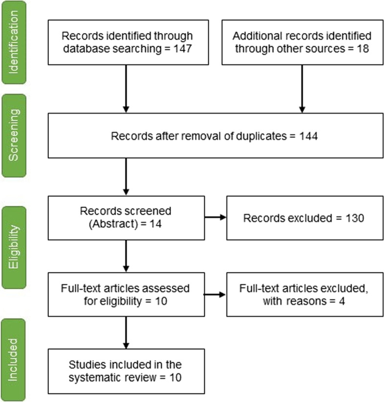
We searched five databases and Google Scholar for articles published between January 2017 and December 2022 that focus on Mycobacterium tuberculosis detection, or tuberculosis pathology using digitised human lung tissue images. Details regarding design, image processing and computer-aided techniques, deep learning models, and datasets were collected and summarised. Discussions, analysis, and comparisons of state-of-the-art methods are provided to help guide future research. Further, a brief update on the relevant techniques and their performance is provided.

RESULTS

Several studies have been conducted to develop automated and AI-assisted methods for diagnosing Mtb and TB pathology from digitised human lung tissue images. Some studies presented a completely automated method of diagnosis, while other studies developed AI-assisted diagnostic methods. Low-level focus areas included the development of a novel CT scanner for soft tissue image contract, and use of multiresolution computed tomography to analyse the 3D structure of the human lung. High-level focus areas included the investigation the effects of aging on the number and size of small airways in the lungs using CT and whole lung high-resolution CT, and the 3D microanatomy characterisation of human tuberculosis lung using CT in conjunction with histology and immunohistochemistry. Additionally, a novel method for acquiring high-resolution 3D images of human lung structure and topology is also presented.

CONCLUSION

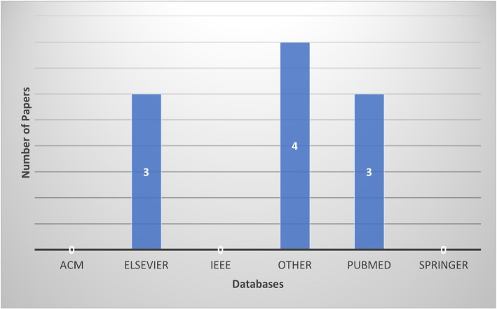
The literature indicates that post 1950s, TB was predominantly studied using animal models even though no animal model reflects the full spectrum of human pulmonary TB disease and does not reproducibly transmit Mtb infection to other animals (Hunter, 2011). This explains why there are very few studies that used human lung tissue for detection or analysis of Mtb. Nonetheless, we found 10 studies that used human tissues (predominately lung) of which five studies proposed machine learning (ML) models for the detection of bacilli and the other five used CT on human lung tissue scanned ex-vivo.

https://cdn.ncbi.nlm.nih.gov/pmc/blobs/61ce/11536899/11573f8191af/12880_2024_1443_Fig3_HTML.jpg
https://cdn.ncbi.nlm.nih.gov/pmc/blobs/61ce/11536899/7d8b7c9b4204/12880_2024_1443_Fig1_HTML.jpg
https://cdn.ncbi.nlm.nih.gov/pmc/blobs/61ce/11536899/3a35f8553538/12880_2024_1443_Fig2_HTML.jpg
https://cdn.ncbi.nlm.nih.gov/pmc/blobs/61ce/11536899/11573f8191af/12880_2024_1443_Fig3_HTML.jpg
https://cdn.ncbi.nlm.nih.gov/pmc/blobs/61ce/11536899/7d8b7c9b4204/12880_2024_1443_Fig1_HTML.jpg
https://cdn.ncbi.nlm.nih.gov/pmc/blobs/61ce/11536899/3a35f8553538/12880_2024_1443_Fig2_HTML.jpg
https://cdn.ncbi.nlm.nih.gov/pmc/blobs/61ce/11536899/11573f8191af/12880_2024_1443_Fig3_HTML.jpg
摘要

目的

对使用数字化人体肺部图像,通过自动或半自动方法检测、诊断或分析结核病(TB)病理学或杆菌的计算机视觉应用进行系统评价。我们将计算机视觉平台分为四类技术:图像处理、目标/模式识别、计算机图形学和深度学习。在本文中,重点是针对 2D 或 3D 数字化人体肺部图像的图像处理和深度学习(DL)应用。这篇综述有助于建立使用人体肺部组织进行 TB 分析的通用实践,并确定该领域进一步研究的机会。该综述提请注意用于检测 TB 的最先进技术,重点介绍当前技术的挑战和局限性。最终目标是促进更高效和准确的 TB 检测或分析算法的发展,并提高对早期检测重要性的认识。

设计

我们在 2017 年 1 月至 2022 年 12 月期间在五个数据库和 Google Scholar 上搜索了重点关注使用数字化人体肺部图像检测结核分枝杆菌或结核病病理学的文章。收集并总结了有关设计、图像处理和计算机辅助技术、深度学习模型和数据集的详细信息。提供了对最先进方法的讨论、分析和比较,以帮助指导未来的研究。此外,还提供了相关技术及其性能的简要更新。

结果

已经进行了多项研究,以开发用于从数字化人体肺部图像诊断 Mtb 和 TB 病理学的自动化和人工智能辅助方法。一些研究提出了一种完全自动化的诊断方法,而其他研究则开发了人工智能辅助诊断方法。低水平重点领域包括开发一种新的软组织 CT 扫描仪来进行软组织图像对比度,以及使用多分辨率 CT 来分析人体肺部的 3D 结构。高水平重点领域包括使用 CT 结合组织学和免疫组织化学研究年龄对肺部小气道数量和大小的影响,以及使用 CT 结合组织学和免疫组织化学对人类肺结核肺的 3D 微观解剖结构进行特征描述。此外,还提出了一种获取人体肺部结构和拓扑的高分辨率 3D 图像的新方法。

结论

文献表明,自 20 世纪 50 年代以来,TB 主要使用动物模型进行研究,尽管没有一种动物模型能反映人类肺部 TB 疾病的全貌,也不能将 Mtb 感染复制性地传播给其他动物(Hunter,2011)。这解释了为什么很少有研究使用人体肺组织来检测或分析 Mtb。尽管如此,我们还是发现了 10 项使用人体组织(主要是肺)的研究,其中 5 项研究提出了用于检测杆菌的机器学习(ML)模型,另外 5 项研究使用了对离体扫描的人体肺部 CT。

相似文献

1
Computer vision applications for the detection or analysis of tuberculosis using digitised human lung tissue images - a systematic review.基于数字化人肺组织图像的计算机视觉技术在结核病检测或分析中的应用:系统综述。
BMC Med Imaging. 2024 Nov 5;24(1):298. doi: 10.1186/s12880-024-01443-w.
2
Computer-aided detection and quantification of cavitary tuberculosis from CT scans.计算机辅助检测和定量分析 CT 扫描中的空洞性肺结核。
Med Phys. 2013 Nov;40(11):113701. doi: 10.1118/1.4824979.
3
Developing and verifying automatic detection of active pulmonary tuberculosis from multi-slice spiral CT images based on deep learning.基于深度学习的多层螺旋 CT 图像活动性肺结核自动检测的建立与验证。
J Xray Sci Technol. 2020;28(5):939-951. doi: 10.3233/XST-200662.
4
Computational techniques for the automated detection of mycobacterium tuberculosis from digitized sputum smear microscopic images: A systematic review.基于数字化痰涂片显微镜图像的结核分枝杆菌自动检测的计算技术:系统评价。
Prog Biophys Mol Biol. 2022 Jul;171:4-16. doi: 10.1016/j.pbiomolbio.2022.03.004. Epub 2022 Mar 23.
5
A fully automatic artificial intelligence-based CT image analysis system for accurate detection, diagnosis, and quantitative severity evaluation of pulmonary tuberculosis.一种基于全自动人工智能的 CT 图像分析系统,用于准确检测、诊断和定量评估肺结核的严重程度。
Eur Radiol. 2022 Apr;32(4):2188-2199. doi: 10.1007/s00330-021-08365-z. Epub 2021 Nov 29.
6
Object recognition in medical images via anatomy-guided deep learning.通过解剖学引导的深度学习实现医学图像中的目标识别。
Med Image Anal. 2022 Oct;81:102527. doi: 10.1016/j.media.2022.102527. Epub 2022 Jun 25.
7
Computed Tomography-Based Biomarker for Longitudinal Assessment of Disease Burden in Pulmonary Tuberculosis.基于计算机断层扫描的生物标志物用于肺结核疾病负担的纵向评估。
Mol Imaging Biol. 2019 Feb;21(1):19-24. doi: 10.1007/s11307-018-1215-x.
8
Automatic discovery of clinically interpretable imaging biomarkers for Mycobacterium tuberculosis supersusceptibility using deep learning.利用深度学习自动发现结核分枝杆菌超敏的临床可解释成像生物标志物。
EBioMedicine. 2020 Dec;62:103094. doi: 10.1016/j.ebiom.2020.103094. Epub 2020 Nov 7.
9
Deep learning techniques in PET/CT imaging: A comprehensive review from sinogram to image space.深度学习技术在 PET/CT 成像中的应用:从能谱到图像空间的全面综述。
Comput Methods Programs Biomed. 2024 Jan;243:107880. doi: 10.1016/j.cmpb.2023.107880. Epub 2023 Oct 21.
10
Folic acid supplementation and malaria susceptibility and severity among people taking antifolate antimalarial drugs in endemic areas.在流行地区,服用抗叶酸抗疟药物的人群中,叶酸补充剂与疟疾易感性和严重程度的关系。
Cochrane Database Syst Rev. 2022 Feb 1;2(2022):CD014217. doi: 10.1002/14651858.CD014217.

本文引用的文献

1
A New Artificial Intelligence-Based Method for Identifying Mycobacterium Tuberculosis in Ziehl-Neelsen Stain on Tissue.一种基于人工智能的在组织齐-尼氏染色中鉴定结核分枝杆菌的新方法。
Diagnostics (Basel). 2022 Jun 17;12(6):1484. doi: 10.3390/diagnostics12061484.
2
A multiscale X-ray phase-contrast tomography dataset of a whole human left lung.一个整个人体左肺的多尺度 X 射线相衬层析数据集。
Sci Data. 2022 Jun 2;9(1):264. doi: 10.1038/s41597-022-01353-y.
3
Deep-Learning-Aided Detection of Mycobacteria in Pathology Specimens Increases the Sensitivity in Early Diagnosis of Pulmonary Tuberculosis Compared with Bacteriology Tests.
与细菌学检测相比,深度学习辅助检测病理标本中的分枝杆菌可提高肺结核早期诊断的敏感性。
Diagnostics (Basel). 2022 Mar 14;12(3):709. doi: 10.3390/diagnostics12030709.
4
Micro-Computed Tomography Analysis of the Human Tuberculous Lung Reveals Remarkable Heterogeneity in Three-dimensional Granuloma Morphology.人体结核性肺部的微计算机断层扫描分析揭示三维肉芽肿形态存在显著异质性。
Am J Respir Crit Care Med. 2021 Sep 1;204(5):583-595. doi: 10.1164/rccm.202101-0032OC.
5
Artificial Intelligence-Based Screening for Mycobacteria in Whole-Slide Images of Tissue Samples.基于人工智能的组织切片全视野图像中分枝杆菌的筛查。
Am J Clin Pathol. 2021 Jun 17;156(1):117-128. doi: 10.1093/ajcp/aqaa215.
6
Small airway loss in the physiologically ageing lung: a cross-sectional study in unused donor lungs.生理老化肺中小气道丢失:未使用供体肺的横断面研究。
Lancet Respir Med. 2021 Feb;9(2):167-174. doi: 10.1016/S2213-2600(20)30324-6. Epub 2020 Oct 5.
7
A CNN-based active learning framework to identify mycobacteria in digitized Ziehl-Neelsen stained human tissues.一种基于卷积神经网络的主动学习框架,用于在数字化齐尔-尼尔森染色的人体组织中识别分枝杆菌。
Comput Med Imaging Graph. 2020 Sep;84:101752. doi: 10.1016/j.compmedimag.2020.101752. Epub 2020 Jul 25.
8
Comprehensive stereological assessment of the human lung using multiresolution computed tomography.使用多分辨率计算机断层扫描对人类肺部进行全面的体视学评估。
J Appl Physiol (1985). 2020 Jun 1;128(6):1604-1616. doi: 10.1152/japplphysiol.00803.2019. Epub 2020 Apr 16.
9
Embracing imperfect datasets: A review of deep learning solutions for medical image segmentation.拥抱不完美数据集:医学图像分割深度学习解决方案综述。
Med Image Anal. 2020 Jul;63:101693. doi: 10.1016/j.media.2020.101693. Epub 2020 Apr 3.
10
Quantitative Assessment of the Effects of Compression on Deep Learning in Digital Pathology Image Analysis.数字病理学图像分析中压缩对深度学习影响的定量评估
JCO Clin Cancer Inform. 2020 Mar;4:221-233. doi: 10.1200/CCI.19.00068.