Department of Radiology, University Hospital, LMU Munich, Munich, Germany.
Clinic for Radiology, University Hospital Muenster, Muenster, Germany.
Cardiovasc Intervent Radiol. 2022 Jul;45(7):992-1000. doi: 10.1007/s00270-022-03169-0. Epub 2022 Jun 2.
To evaluate the safety and outcome of image-guided embolotherapy of extracranial arteriovenous malformations (AVMs) primarily affecting the face.
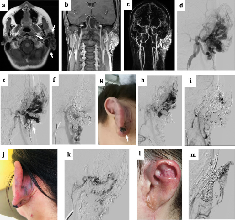
A multicenter cohort of 28 patients presenting with AVMs primarily affecting the face was retrospectively investigated. Fifty image-guided embolotherapies were performed, mostly using ethylene-vinyl alcohol copolymer-based embolic agents. Clinical and imaging findings were assessed to evaluate response during follow-up (symptom-free, partial relief of symptoms, no improvement, and progression despite embolization), lesion devascularization (total, 100%; substantial, 76-99%; partial, 51-75%; failure, < 50%; and progression), and complication rates (classified according to the CIRSE guidelines). Sub-analyses regarding clinical outcome (n = 24) were performed comparing patients with (n = 12) or without (n = 12) subsequent surgical resection after embolotherapy.
The median number of embolotherapy sessions was 2.0 (range, 1-4). Clinical outcome after a mean follow-up of 12.4 months (± 13.3; n = 24) revealed a therapy response in 21/24 patients (87.5%). Imaging showed total devascularization in 14/24 patients (58.3%), including the 12 patients with subsequent surgery and 2 additional patients with embolotherapy only. Substantial devascularization (76-99%) was assessed in 7/24 patients (29.2%), and partial devascularization (51-75%) in 3/24 patients (12.5%). Complications occurred during/after 12/50 procedures (24.0%), including 18.0% major complications. Patients with subsequent surgical resections were more often symptom-free at the last follow-up compared to the group having undergone embolotherapy only (p = 0.006).
Image-guided embolotherapy is safe and effective for treating extracranial AVMs of the face. Subsequent surgical resections after embolization may substantially improve patients' clinical outcome, emphasizing the need for multimodal therapeutic concepts.
Level 4, Retrospective study.
评估主要影响面部的颅外动静脉畸形(AVM)的图像引导栓塞治疗的安全性和结果。
回顾性调查了 28 例主要影响面部的 AVM 患者的多中心队列。进行了 50 次图像引导栓塞治疗,主要使用乙烯-乙烯醇共聚物基栓塞剂。评估临床和影像学发现,以在随访期间评估反应(无症状、症状部分缓解、无改善和栓塞后进展)、病变血管化程度(完全,100%;显著,76-99%;部分,51-75%;失败,<50%;进展)和并发症发生率(根据 CIRSE 指南分类)。对栓塞治疗后行(n=12)或不行(n=12)后续手术切除的患者进行了临床结局(n=24)的亚分析比较。
中位数栓塞治疗次数为 2.0(范围,1-4)。平均 12.4 个月(±13.3;n=24)的随访后临床结局显示 24 例患者中有 21 例(87.5%)有治疗反应。影像学显示 14 例患者(58.3%)完全血管化,其中 12 例患者行后续手术,2 例患者仅行栓塞治疗。7 例患者(29.2%)评估为显著血管化(76-99%),3 例患者(12.5%)为部分血管化(51-75%)。50 次手术中有 12 次(24.0%)术中/术后发生并发症,包括 18.0%的严重并发症。最后一次随访时,行后续手术切除的患者无症状的比例高于仅行栓塞治疗的患者(p=0.006)。
图像引导栓塞治疗是治疗面部颅外 AVM 的安全有效的方法。栓塞后行后续手术切除可能会显著改善患者的临床结局,强调需要采用多模态治疗方案。
4 级,回顾性研究。