Department of Internal Medicine and Oncology, Faculty of Medicine, Semmelweis University, 1083, Budapest, Hungary.
Molecular Medicine Research Group, Eötvös Loránd Research Network, 1083, Budapest, Hungary.
BMC Cancer. 2022 Jun 2;22(1):605. doi: 10.1186/s12885-022-09659-1.
Hypomethylation of long interspersed nuclear element 1 (LINE-1) is characteristic of various cancer types, including colorectal cancer (CRC). Malfunction of several factors or alteration of methyl-donor molecules' (folic acid and S-adenosylmethionine) availability can contribute to DNA methylation changes. Detection of epigenetic alterations in liquid biopsies can assist in the early recognition of CRC. Following the investigations of a Hungarian colon tissue sample set, our goal was to examine the LINE-1 methylation of blood samples along the colorectal adenoma-carcinoma sequence and in inflammatory bowel disease. Moreover, we aimed to explore the possible underlying mechanisms of global DNA hypomethylation formation on a multi-level aspect.
LINE-1 methylation of colon tissue (n = 183) and plasma (n = 48) samples of healthy controls and patients with colorectal tumours were examined with bisulfite pyrosequencing. To investigate mRNA expression, microarray analysis results were reanalysed in silico (n = 60). Immunohistochemistry staining was used to validate DNA methyltransferases (DNMTs) and folate receptor beta (FOLR2) expression along with the determination of methyl-donor molecules' in situ level (n = 40).
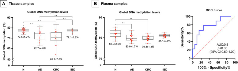
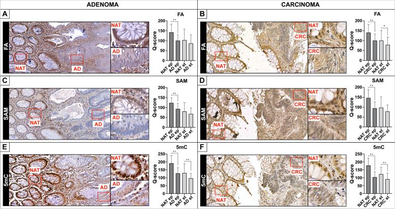
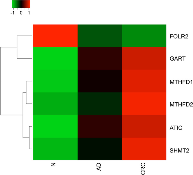
Significantly decreased LINE-1 methylation level was observed in line with cancer progression both in tissue (adenoma: 72.7 ± 4.8%, and CRC: 69.7 ± 7.6% vs. normal: 77.5 ± 1.7%, p ≤ 0.01) and liquid biopsies (adenoma: 80.0 ± 1.7%, and CRC: 79.8 ± 1.3% vs. normal: 82.0 ± 2.0%, p ≤ 0.01). However, no significant changes were recognized in inflammatory bowel disease cases. According to in silico analysis of microarray data, altered mRNA levels of several DNA methylation-related enzymes were detected in tumours vs. healthy biopsies, namely one-carbon metabolism-related genes-which met our analysing criteria-showed upregulation, while FOLR2 was downregulated. Using immunohistochemistry, DNMTs, and FOLR2 expression were confirmed. Moreover, significantly diminished folic acid and S-adenosylmethionine levels were observed in parallel with decreasing 5-methylcytosine staining in tumours compared to normal adjacent to tumour tissues (p ≤ 0.05).
Our results suggest that LINE-1 hypomethylation may have a distinguishing value in precancerous stages compared to healthy samples in liquid biopsies. Furthermore, the reduction of global DNA methylation level could be linked to reduced methyl-donor availability with the contribution of decreased FOLR2 expression.
长散布核元件 1(LINE-1)的低甲基化是包括结直肠癌(CRC)在内的多种癌症类型的特征。几种因素的功能障碍或甲基供体分子(叶酸和 S-腺苷甲硫氨酸)可用性的改变可能导致 DNA 甲基化的改变。液体活检中表观遗传改变的检测有助于早期识别 CRC。在对一组匈牙利结肠组织样本进行调查后,我们的目标是检查血液样本中 LINE-1 的甲基化情况,包括结直肠腺瘤-癌序列和炎症性肠病中的情况。此外,我们旨在从多个层面探索形成全基因组低甲基化的可能潜在机制。
使用亚硫酸氢盐焦磷酸测序法检测健康对照组和结直肠肿瘤患者的结肠组织(n=183)和血浆(n=48)样本中的 LINE-1 甲基化情况。为了研究 mRNA 表达,我们对微阵列分析的结果进行了计算机分析(n=60)。免疫组织化学染色用于验证 DNA 甲基转移酶(DNMTs)和叶酸受体β(FOLR2)的表达情况,并同时确定甲基供体分子的原位水平(n=40)。
我们观察到,组织(腺瘤:72.7±4.8%,CRC:69.7±7.6%,与正常组织:77.5±1.7%,p≤0.01)和液体活检(腺瘤:80.0±1.7%,CRC:79.8±1.3%,与正常组织:82.0±2.0%,p≤0.01)中 LINE-1 甲基化水平随着癌症的进展而显著降低。然而,在炎症性肠病病例中没有观察到显著变化。根据微阵列数据的计算机分析,与健康活检相比,肿瘤中几种 DNA 甲基化相关酶的 mRNA 水平发生了改变,符合我们分析标准的一碳代谢相关基因上调,而 FOLR2 下调。通过免疫组织化学染色,证实了 DNMTs 和 FOLR2 的表达。此外,与肿瘤旁正常组织相比,肿瘤中观察到叶酸和 S-腺苷甲硫氨酸水平显著降低,同时 5-甲基胞嘧啶染色减少(p≤0.05)。
我们的研究结果表明,与健康样本相比,LINE-1 低甲基化在液体活检的癌前阶段可能具有鉴别价值。此外,全基因组 DNA 甲基化水平的降低可能与甲基供体可用性的降低有关,这可能与 FOLR2 表达的降低有关。