Stolz Bernadette J, Kaeppler Jakob, Markelc Bostjan, Braun Franziska, Lipsmeier Florian, Muschel Ruth J, Byrne Helen M, Harrington Heather A
Mathematical Institute, University of Oxford, Oxford, UK.
Oxford Institute for Radiation Oncology, University of Oxford, Oxford, UK.
Sci Adv. 2022 Jun 10;8(23):eabm2456. doi: 10.1126/sciadv.abm2456.
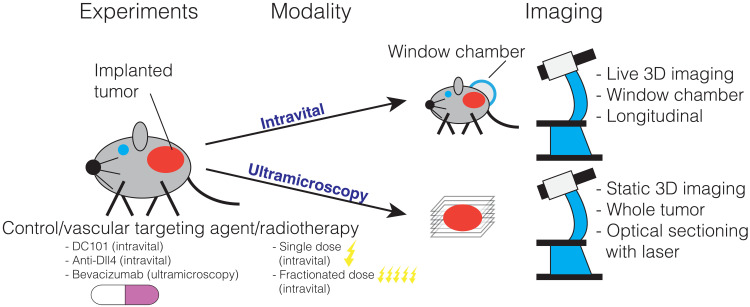
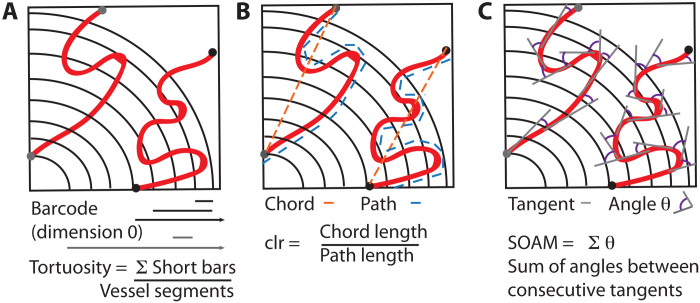
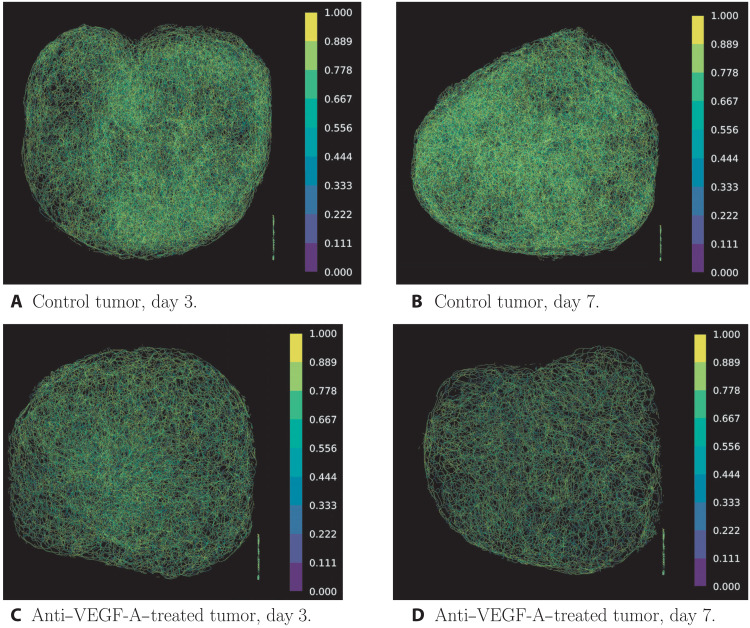
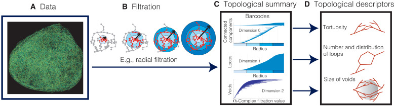
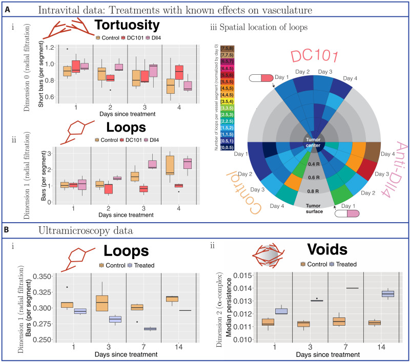
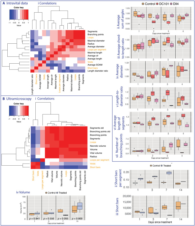
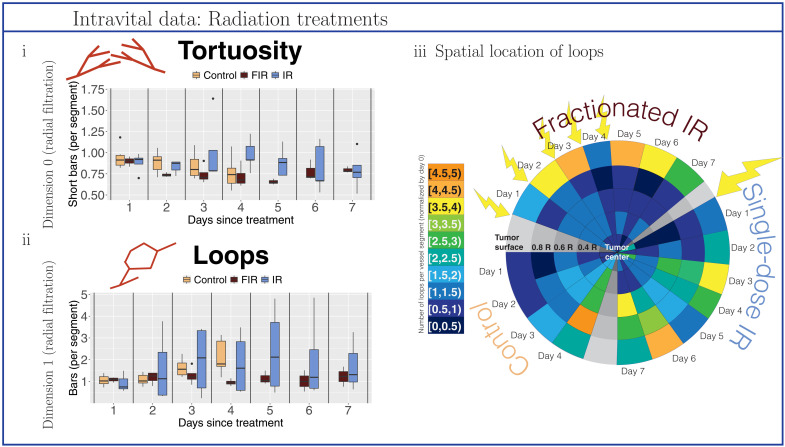
Advances in imaging techniques enable high-resolution three-dimensional (3D) visualization of vascular networks over time and reveal abnormal structural features such as twists and loops, and their quantification is an active area of research. Here, we showcase how topological data analysis, the mathematical field that studies the "shape" of data, can characterize the geometric, spatial, and temporal organization of vascular networks. We propose two topological lenses to study vasculature, which capture inherent multiscale features and vessel connectivity, and surpass the single-scale analysis of existing methods. We analyze images collected using intravital and ultramicroscopy modalities and quantify spatiotemporal variation of twists, loops, and avascular regions (voids) in 3D vascular networks. This topological approach validates and quantifies known qualitative trends such as dynamic changes in tortuosity and loops in response to antibodies that modulate vessel sprouting; furthermore, it quantifies the effect of radiotherapy on vessel architecture.
成像技术的进步使得能够随时间对血管网络进行高分辨率三维(3D)可视化,并揭示诸如扭曲和环圈等异常结构特征,而对其进行量化是一个活跃的研究领域。在此,我们展示了拓扑数据分析这一研究数据“形状”的数学领域如何能够表征血管网络的几何、空间和时间组织。我们提出了两种拓扑视角来研究脉管系统,它们能够捕捉固有的多尺度特征和血管连通性,并且超越了现有方法的单尺度分析。我们分析了使用活体成像和超显微镜成像模式收集的图像,并量化了三维血管网络中扭曲、环圈和无血管区域(空隙)的时空变化。这种拓扑方法验证并量化了已知的定性趋势,例如响应调节血管生成的抗体时曲折度和环圈的动态变化;此外,它还量化了放射疗法对血管结构的影响。