Department of Orthopaedic, Trauma, and Reconstructive Surgery, RWTH University Hospital, Pauwelsstraße 30, 52074, Aachen, Germany.
Department of Ophthalmology, RWTH University Hospital, Pauwelsstr. 30, 52074, Aachen, Germany.
J Orthop Surg Res. 2022 Jun 11;17(1):311. doi: 10.1186/s13018-022-03201-6.
Some authors secured the membrane during matrix-induced autologous chondrocyte implantation (mACI) with fibrin glue or did not use a formal fixation. The real impact of fibrin glue addition on chondrocytes migration and proliferation has not yet been clarified. This study evaluated the impact of fibrin glue on a chondrocyte loaded collagenic membrane.
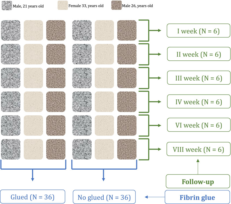
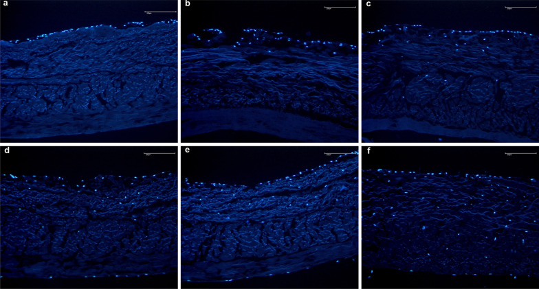
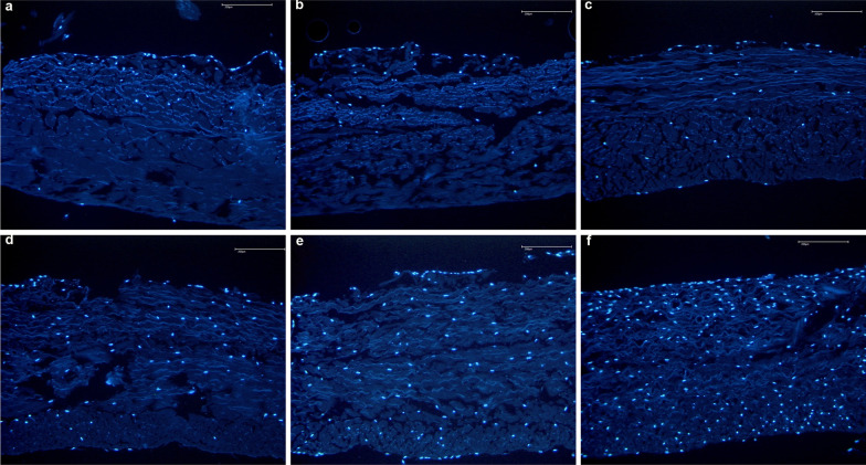
A resorbable collagen I/III porcine derived membrane commonly employed in AMIC was used for all experiments. Chondrocytes from three difference donors were used. At 1-, 2-, 3-, 4-, 6-, and at 8-week the membranes were embedded in Mounting Medium with Dapi (ABCAM, Cambridge, UK). The Dapi contained in the mounting medium ties the DNA of the cell nucleus and emits a blue fluorescence. In this way, the spreading of the cells in the membrane can be easily monitored. The outcomes of interest were to evaluate (1) cell migration and (2) cell proliferation within the porous membrane layer. DAPI/nuclei signals were analysed with fluorescence microscope under a magnification of 100-fold.
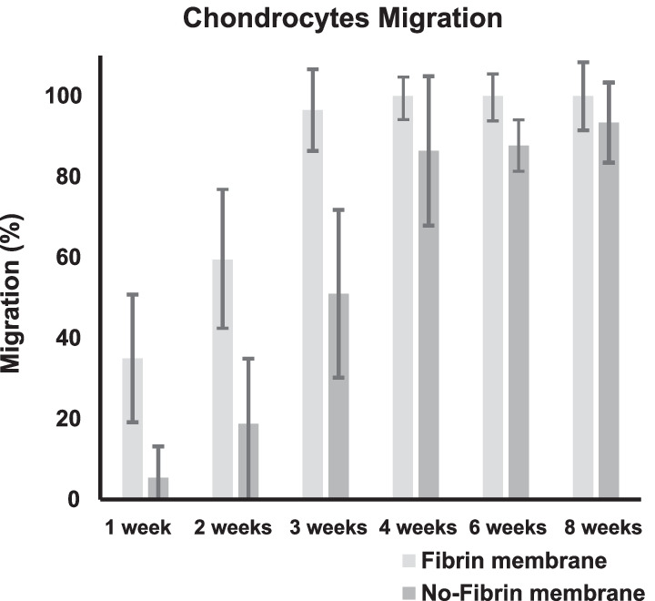
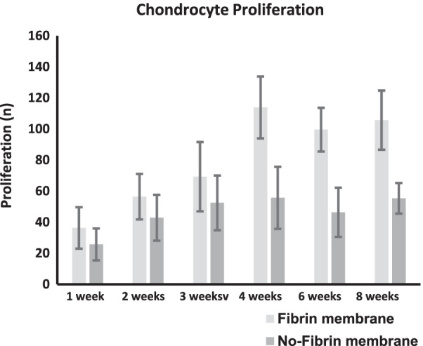
The no-fibrin group demonstrated greater migration of the cells within the membrane. Although migration resulted higher in the no-fibrin group at every follow-up, this difference was significant only at week 1 (P < 0.001), 2 (P = 0.004), and 3 (P = 0.03). No difference was found at week 3, 6, and 8. The no-fibrin group demonstrated greater proliferation of the chondrocytes within the membrane. These differences were significant at week 4 (P < 0.0001), 6 (P < 0.0001), 8 (P < 0.0001).
The use of fibrin glue over a resorbable membrane leads to lower in vitro proliferation and migration of chondrocytes.
一些作者在基质诱导的自体软骨细胞植入术(mACI)中使用纤维蛋白胶固定膜,或者不进行正式固定。纤维蛋白胶的添加对软骨细胞迁移和增殖的实际影响尚未阐明。本研究评估了纤维蛋白胶对负载软骨细胞的胶原膜的影响。
所有实验均使用 AMIC 中常用的可吸收胶原 I/III 猪衍生膜。使用来自三个不同供体的软骨细胞。在第 1、2、3、4、6 和 8 周时,将膜嵌入含 Dapi 的 Mounting Medium(ABCAM,英国剑桥)中。Mounting Medium 中含有的 Dapi 与细胞核的 DNA 结合并发出蓝色荧光。通过这种方式,可以轻松监测细胞在膜中的扩散。研究的重点是评估(1)细胞在多孔膜层内的迁移和(2)细胞增殖。用荧光显微镜在 100 倍放大倍数下分析 DAPI/细胞核信号。
无纤维蛋白组的细胞在膜内的迁移率更高。虽然无纤维蛋白组在每个随访时间点的迁移率均较高,但仅在第 1 周(P<0.001)、第 2 周(P=0.004)和第 3 周(P=0.03)差异有统计学意义。在第 3、6 和 8 周时未发现差异。无纤维蛋白组的细胞在膜内的增殖率更高。这些差异在第 4 周(P<0.0001)、第 6 周(P<0.0001)和第 8 周(P<0.0001)时具有统计学意义。
在可吸收膜上使用纤维蛋白胶会导致软骨细胞体外增殖和迁移率降低。