Liang Rong-Bin, Liu Li-Qi, Shi Wen-Qing, Sun Tie, Ge Qian-Min, Li Qiu-Yu, Shu Hui-Ye, Zhang Li-Juan, Shao Yi
Department of Ophthalmology, The First Affiliated Hospital of Nanchang University, Jiangxi Center of National Ocular Disease Clinical Research, Nanchang, China.
Front Hum Neurosci. 2022 May 24;16:900409. doi: 10.3389/fnhum.2022.900409. eCollection 2022.
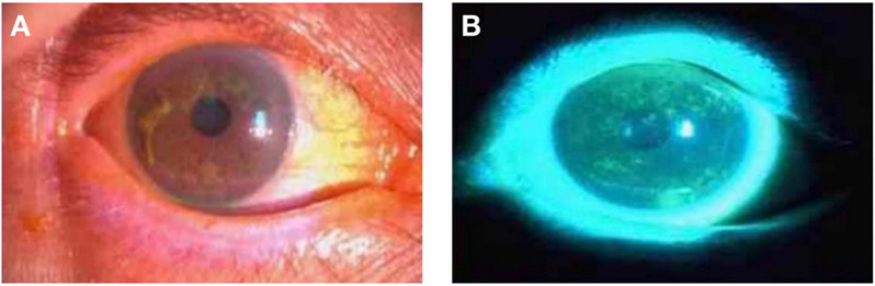
To investigate spontaneous brain activity in patients with dry eye (DE) and healthy control (HC) using the fractional amplitude of low frequency fluctuation (fALFF) technique with the aim of elucidating the relationship between the clinical symptoms of DE and changes in brain function.
A total of 28 patients with DE and 28 matched healthy volunteers (10 males and 18 females in each group) were enrolled. Resting-state functional magnetic resonance imaging scans were performed in both groups. Then all subjects were required to complete a comprehensive Hospital Anxiety and Depression Scale (HADS). Receiver operating characteristic (ROC) curve analysis was used to evaluate the differences in fALFF values between the two groups and their diagnostic value. Linear correlations between HADS and fALFF values in different brain regions of DE patients were analyzed using the correlation coefficient.
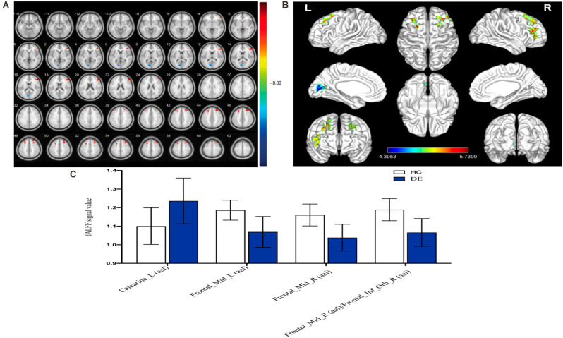
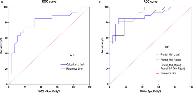
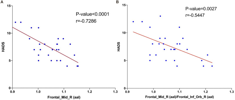
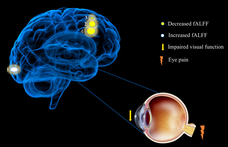
Patients with DE had significantly higher fALFF values in the left calcarine sulcus (CS) than the HC group, while fALFF values in the bilateral middle frontal gyrus (MFG) and right MFG/right inferior frontal gyrus (IFG) were significantly lower in DE patients than in HC group. fALFF values had a high diagnostic value for differentiating patients with DE from the HC group ( < 0.001). Right MFG and right MFG/IFG were significantly correlated with HADS values.
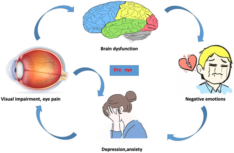
Our study found that DE mainly involved functional disorders in the brain areas of the left CS, bilateral MFG and right MFG/right IFG, which helped us to find possible clinical features of DE disease and reflected the potential pathological mechanism of DE.
采用低频振幅分数(fALFF)技术研究干眼症(DE)患者和健康对照者(HC)的自发脑活动,以阐明DE临床症状与脑功能变化之间的关系。
共纳入28例DE患者和28例匹配的健康志愿者(每组10例男性和18例女性)。两组均进行静息态功能磁共振成像扫描。然后所有受试者均需完成一份全面的医院焦虑抑郁量表(HADS)。采用受试者工作特征(ROC)曲线分析评估两组间fALFF值的差异及其诊断价值。使用相关系数分析DE患者不同脑区HADS与fALFF值之间的线性相关性。
DE患者左侧距状沟(CS)的fALFF值显著高于HC组,而DE患者双侧额中回(MFG)和右侧MFG/右侧额下回(IFG)的fALFF值显著低于HC组。fALFF值对区分DE患者和HC组具有较高的诊断价值(<0.001)。右侧MFG和右侧MFG/IFG与HADS值显著相关。
我们的研究发现,DE主要涉及左侧CS、双侧MFG和右侧MFG/右侧IFG脑区的功能障碍,这有助于我们发现DE疾病可能的临床特征,并反映了DE的潜在病理机制。