Chin Brian W, King Kassandra A, George Nicholas K, Neeki Michael M, Mistry Jamshid T
Arrowhead Regional Medical Center, Department of Emergency Medicine, Colton, California.
Arrowhead Regional Medical Center, Department of Medicine, Colton, California.
Clin Pract Cases Emerg Med. 2022 May;6(2):1-4. doi: 10.5811/cpcem.2021.9.53553.
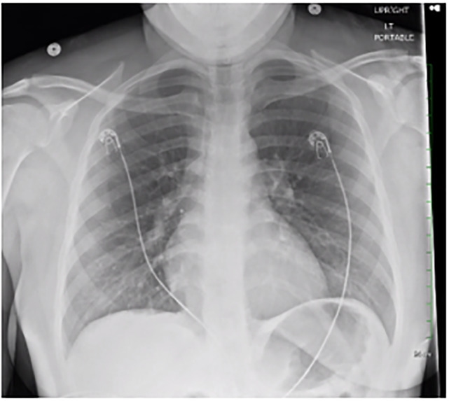
Cardiac masses are a rare cause of chest pain. They can often be missed on a chest radiograph performed to evaluate non-specific chest pain and are not readily evaluated with traditional laboratory testing. However, these masses can be visualized with point-of-care ultrasound.
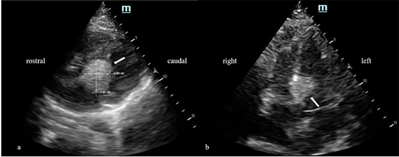
We present a case of a 19-year-old female presenting with intermittent chest pain, palpitations, and weakness present for two months. The patient had previously been evaluated at our emergency department one week earlier and was diagnosed with anxiety before being discharged. Besides a tachycardic and labile heart rate, physical examination and laboratory testing were unremarkable. Point-of-care cardiac echocardiography subsequently demonstrated findings concerning for a cardiac mass.
Cardiac masses are a rare cause of chest pain and palpitations that are easily missed. The advent of point-of-care ultrasonography has afforded us the ability to rapidly assess for structural and functional cardiac abnormalities at bedside, and incorporation of this tool into the evaluation of patients with chest pain offers the ability to detect these rare pathologies.
心脏肿物是胸痛的罕见病因。在为评估非特异性胸痛而进行的胸部X光检查中,它们常常会被漏诊,并且传统实验室检查也难以对其进行评估。然而,这些肿物可以通过床旁超声检查显示出来。
我们报告一例19岁女性病例,该患者出现间歇性胸痛、心悸及乏力症状达两个月。患者一周前曾在我们的急诊科就诊,出院前被诊断为焦虑症。除心率过速且不稳定外,体格检查和实验室检查均无异常。随后床旁心脏超声心动图检查显示有心脏肿物的相关表现。
心脏肿物是胸痛和心悸的罕见病因,容易被漏诊。床旁超声检查的出现使我们能够在床边快速评估心脏的结构和功能异常,将这一工具纳入胸痛患者的评估中有助于发现这些罕见病症。