Bashit Abdullah Al, Nepal Prakash, Connors Theresa, Oakley Derek H, Hyman Bradley T, Yang Lin, Makowski Lee
Department of Electrical and Computer Engineering, Northeastern University, Boston, MA, United States.
Department of Bioengineering, Northeastern University, Boston, MA, United States.
Front Neurosci. 2022 Jun 1;16:909542. doi: 10.3389/fnins.2022.909542. eCollection 2022.
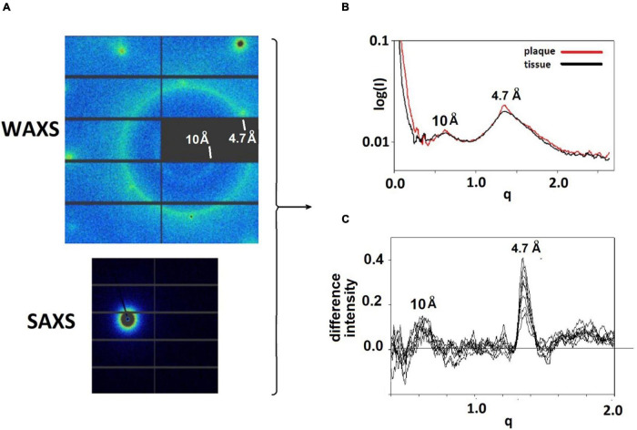
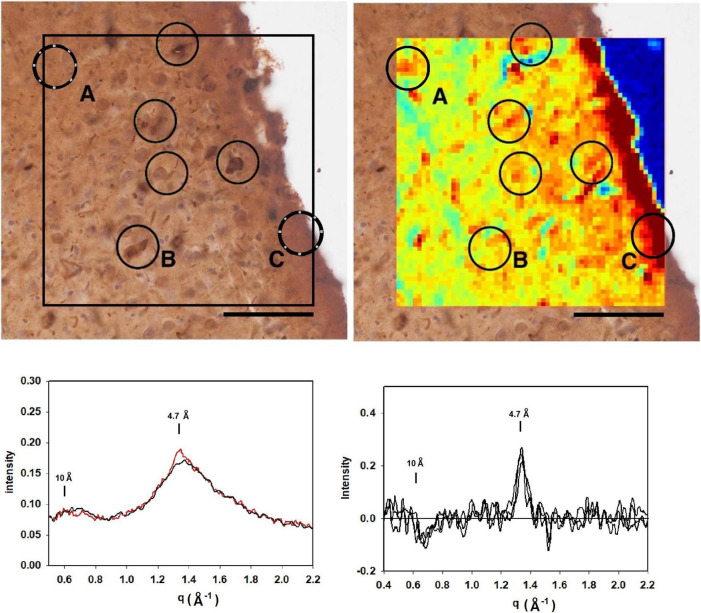
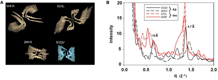
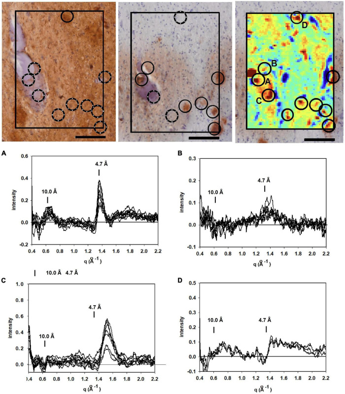
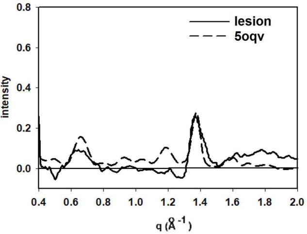
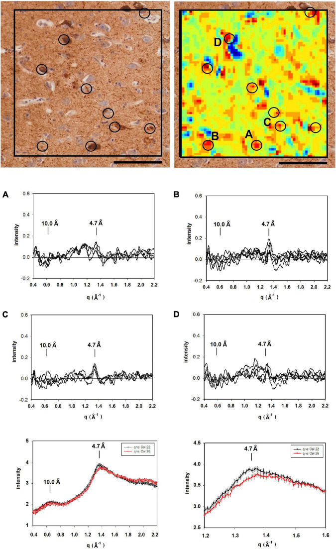
Alzheimer's disease (AD) is a neurodegenerative disorder defined by the progressive formation and spread of fibrillar aggregates of Aβ peptide and tau protein. Polymorphic forms of these aggregates may contribute to disease in varying ways since different neuropathologies appear to be associated with different sets of fibrillar structures and follow distinct pathological trajectories that elicit characteristic clinical phenotypes. The molecular mechanisms underlying the spread of these aggregates in disease may include nucleation, replication, and migration all of which could vary with polymorphic form, stage of disease, and region of brain. Given the linkage between mechanisms of progression and distribution of polymorphs, mapping the distribution of fibrillar structures has the potential to discriminate between mechanisms of progression. However, the means of carrying out this mapping are limited. Optical microscopy lacks the resolution to discriminate between polymorphs , and higher resolution tools such as ssNMR and cryoEM require the isolation of fibrils from tissue, destroying relevant spatial information. Here, we demonstrate the use of scanning x-ray microdiffraction (XMD) to map the locations of fibrillar polymorphs of Aβ peptides and tau protein in histological thin sections of human brain tissue. Coordinated examination of serial sections by immunohistochemistry was used to aid in the interpretation of scattering patterns and to put the observations in a broader anatomical context. Scattering from lesions in tissue shown to be rich in Aβ fibrils by immunohistochemistry exhibited scattering patterns with a prototypical 4.7 Å cross-β peak, and overall intensity distribution that compared well with that predicted from high resolution structures. Scattering from lesions in tissue with extensive tau pathology also exhibited a 4.7 Å cross-β peak but with intensity distributions that were distinct from those seen in Aβ-rich regions. In summary, these observations demonstrate that XMD is a rich source of information on the distribution of fibrillar polymorphs in diseased human brain tissue. When used in coordination with neuropathological examination it has the potential to provide novel insights into the molecular mechanisms underlying disease.
阿尔茨海默病(AD)是一种神经退行性疾病,其特征是β淀粉样蛋白(Aβ)肽和tau蛋白的纤维状聚集体逐渐形成并扩散。这些聚集体的多态形式可能以不同方式导致疾病,因为不同的神经病理学似乎与不同的纤维状结构组相关,并遵循不同的病理轨迹,引发特征性的临床表型。这些聚集体在疾病中扩散的分子机制可能包括成核、复制和迁移,所有这些都可能因多态形式、疾病阶段和脑区而异。鉴于多态体的进展机制与分布之间的联系,绘制纤维状结构的分布图有可能区分进展机制。然而,进行这种绘图的方法有限。光学显微镜缺乏区分多态体的分辨率,而诸如固态核磁共振(ssNMR)和冷冻电子显微镜(cryoEM)等更高分辨率的工具需要从组织中分离纤维,从而破坏相关的空间信息。在这里,我们展示了使用扫描X射线微衍射(XMD)来绘制人脑组织组织学薄切片中Aβ肽和tau蛋白纤维状多态体的位置。通过免疫组织化学对连续切片进行协同检查,以帮助解释散射模式,并将观察结果置于更广泛的解剖背景中。免疫组织化学显示富含Aβ纤维的组织病变的散射呈现出具有典型4.7 Å交叉β峰的散射模式,其整体强度分布与从高分辨率结构预测的结果相当。具有广泛tau病理学的组织病变的散射也呈现出4.7 Å交叉β峰,但强度分布与富含Aβ的区域不同。总之,这些观察结果表明,XMD是关于患病人类脑组织中纤维状多态体分布的丰富信息来源。当与神经病理学检查配合使用时,它有可能为疾病背后的分子机制提供新的见解。