• 文献检索
  • 文档翻译
  • 深度研究
  • 学术资讯
  • Suppr Zotero 插件Zotero 插件
  • 邀请有礼
  • 套餐&价格
  • 历史记录
应用&插件
Suppr Zotero 插件Zotero 插件浏览器插件Mac 客户端Windows 客户端微信小程序
定价
高级版会员购买积分包购买API积分包
服务
文献检索文档翻译深度研究API 文档MCP 服务
关于我们
关于 Suppr公司介绍联系我们用户协议隐私条款
关注我们

Suppr 超能文献

核心技术专利:CN118964589B侵权必究
粤ICP备2023148730 号-1Suppr @ 2026

文献检索

告别复杂PubMed语法,用中文像聊天一样搜索,搜遍4000万医学文献。AI智能推荐,让科研检索更轻松。

立即免费搜索

文件翻译

保留排版,准确专业,支持PDF/Word/PPT等文件格式,支持 12+语言互译。

免费翻译文档

深度研究

AI帮你快速写综述,25分钟生成高质量综述,智能提取关键信息,辅助科研写作。

立即免费体验

靛胭脂增强放大内镜在非壶腹性十二指肠浅表肿瘤中的应用:一项初步研究。

Magnified endoscopy with texture and color enhanced imaging with indigo carmine for superficial nonampullary duodenal tumor: a pilot study.

机构信息

Department of Gastroenterology, Graduate School of Medicine, Chiba University, Inohana 1-8-1, Chiba-City, 260-8670, Japan.

Kameido Endoscopy and Gastroenterology Clinic, Tokyo, Japan.

出版信息

Sci Rep. 2022 Jun 20;12(1):10381. doi: 10.1038/s41598-022-14476-4.

DOI:10.1038/s41598-022-14476-4
PMID:35725752
原文链接:https://pmc.ncbi.nlm.nih.gov/articles/PMC9209444/
Abstract

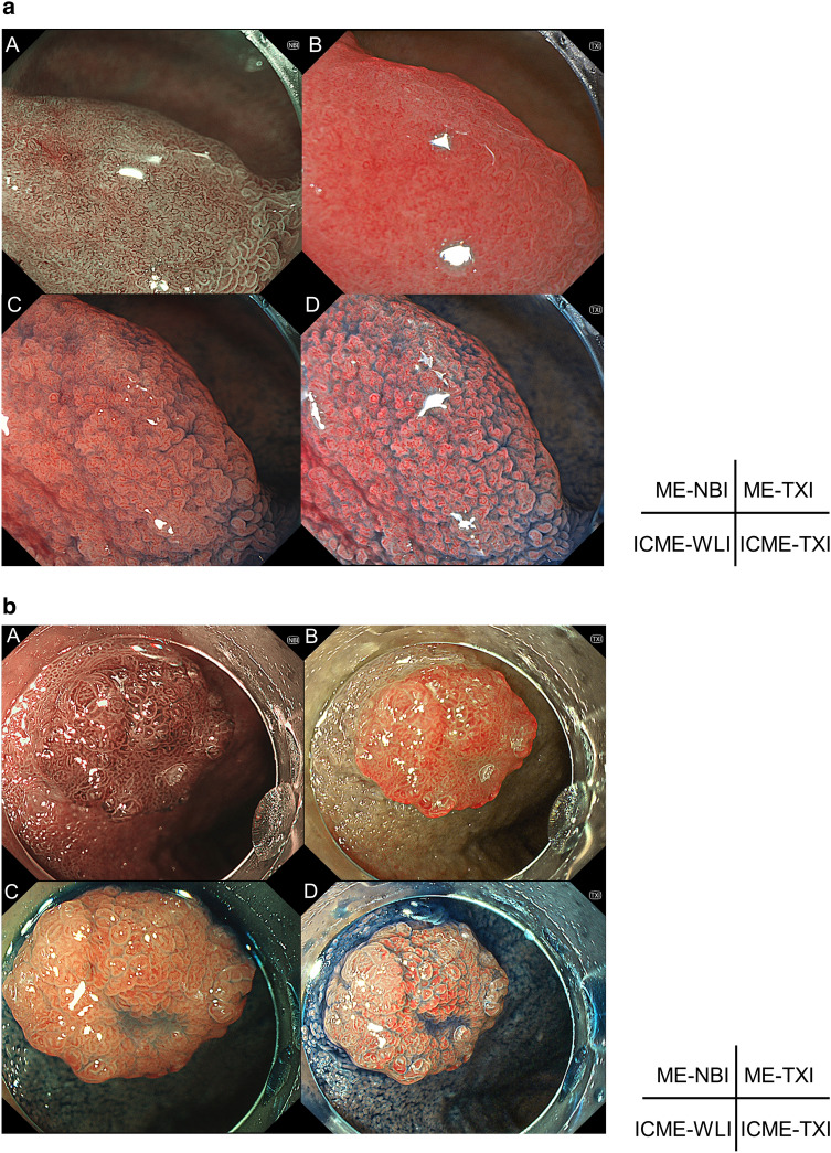
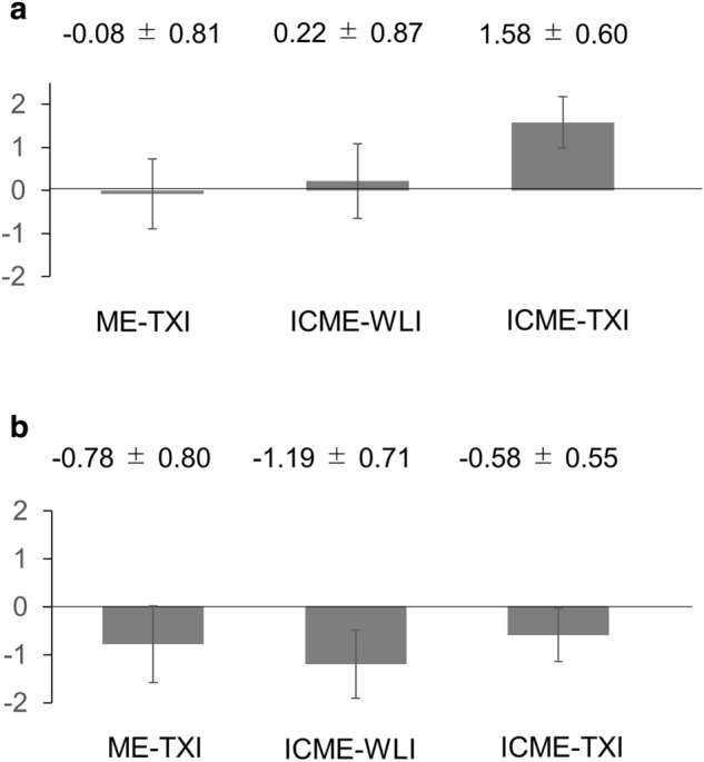
This pilot study aimed to investigate the utility of texture and color enhancement imaging (TXI) with magnified endoscopy (ME) for the preoperative diagnosis of superficial nonampullary duodenal epithelial tumors (SNADETs). We prospectively evaluated 12 SNADETs. The visibility for ME-TXI, ME with indigo carmine (ICME)-white-light imaging (WLI), ICME-TXI compared to ME-NBI (narrow-band imaging) was scored (+ 2 to - 2 ME-NBI was set as score 0) by 3 experts. Scores + 2 and + 1 were defined as improved visibility. The intra-observer and interobserver agreement for improved visibility of surface structure (SS) was evaluated. Sensitivity, specificity, and positive predictive value (PPV) for Vienna Classification (VCL) C4/5 associated with the preoperative diagnosis of ICME-TXI were analyzed. The SS visibility score of ICME-TXI was significantly higher than that of ME-NBI, ME-TXI, and ICME-WLI (P < 0.001 respectively). The kappa coefficients of reliability for intra-observer and interobserver agreement for the SS visibility improvement with ICME-TXI were 0.96, 1.00, 1.00 and 0.70, 0.96, 0.96 respectively. All endoscopists preferred ICME-TXI for visualizing SS mostly for all lesions. The sensitivity, specificity, and PPV (%) of ICME-TXI for VCL C4/5 were 80, 66.7, and 63.2, respectively. ICME-TXI facilitates the visibility of the SS of SNADETs and may contribute to their preoperative diagnosis.

摘要

本研究旨在探讨放大内镜(ME)联合 TXI 对非壶腹型十二指肠上皮性肿瘤(SNADETs)术前诊断的应用价值。我们前瞻性评估了 12 例 SNADETs。3 位专家对 ME-TXI、ICME-WLI、ICME-TXI 与 ME-NBI(窄带成像)的可视性进行评分(+2 至-2,ME-NBI 为 0 分)。评分+2 和+1 定义为可视性改善。评估观察者内和观察者间对表面结构(SS)可视性改善的一致性。分析与术前 ICME-TXI 诊断相关的维也纳分类(VCL)C4/5 的敏感性、特异性和阳性预测值(PPV)。ICME-TXI 的 SS 可视性评分明显高于 ME-NBI、ME-TXI 和 ICME-WLI(P 均<0.001)。ICME-TXI 的 SS 可视性改善的观察者内和观察者间一致性的kappa 系数分别为 0.96、1.00、1.00 和 0.70、0.96、0.96。所有内镜医生均首选 ICME-TXI 观察 SS,主要用于所有病变。ICME-TXI 对 VCL C4/5 的敏感性、特异性和 PPV(%)分别为 80、66.7 和 63.2。ICME-TXI 可改善 SNADETs 的 SS 可视性,有助于其术前诊断。

https://cdn.ncbi.nlm.nih.gov/pmc/blobs/b312/9209444/871ee246c83b/41598_2022_14476_Fig2_HTML.jpg
https://cdn.ncbi.nlm.nih.gov/pmc/blobs/b312/9209444/78de4b73f054/41598_2022_14476_Fig1_HTML.jpg
https://cdn.ncbi.nlm.nih.gov/pmc/blobs/b312/9209444/871ee246c83b/41598_2022_14476_Fig2_HTML.jpg
https://cdn.ncbi.nlm.nih.gov/pmc/blobs/b312/9209444/78de4b73f054/41598_2022_14476_Fig1_HTML.jpg
https://cdn.ncbi.nlm.nih.gov/pmc/blobs/b312/9209444/871ee246c83b/41598_2022_14476_Fig2_HTML.jpg

相似文献

1
Magnified endoscopy with texture and color enhanced imaging with indigo carmine for superficial nonampullary duodenal tumor: a pilot study.靛胭脂增强放大内镜在非壶腹性十二指肠浅表肿瘤中的应用:一项初步研究。
Sci Rep. 2022 Jun 20;12(1):10381. doi: 10.1038/s41598-022-14476-4.
2
Improved visibility of colorectal tumor by texture and color enhancement imaging with indigo carmine.通过靛胭脂纹理和颜色增强成像提高结直肠肿瘤的可视性。
World J Gastrointest Endosc. 2023 Dec 16;15(12):690-698. doi: 10.4253/wjge.v15.i12.690.
3
Texture and color enhancement imaging in magnifying endoscopic evaluation of colorectal adenomas.放大内镜评估大肠腺瘤中的纹理和颜色增强成像
World J Gastrointest Endosc. 2022 Feb 16;14(2):96-105. doi: 10.4253/wjge.v14.i2.96.
4
Analysis of Texture and Color Enhancement Imaging for Improving the Visibility of Non-polypoid Colorectal Lesions.分析纹理和颜色增强成像以提高非息肉样结直肠病变的可视性。
Dig Dis Sci. 2022 Dec;67(12):5657-5665. doi: 10.1007/s10620-022-07460-5. Epub 2022 Mar 22.
5
Comparison of Texture and Color Enhancement Imaging with White Light Imaging in 52 Patients with Short-Segment Barrett's Esophagus.52 例短节段 Barrett 食管患者的纹理及色彩增强成像与白光成像的对比。
Med Sci Monit. 2023 Jun 13;29:e940249. doi: 10.12659/MSM.940249.
6
Texture and Color Enhancement Imaging Increases Color Changes and Improves Visibility for Squamous Cell Carcinoma Suspicious Lesions in the Pharynx and Esophagus.纹理与颜色增强成像增加了颜色变化并提高了咽部和食管鳞状细胞癌可疑病变的可视性。
Diagnostics (Basel). 2021 Oct 23;11(11):1971. doi: 10.3390/diagnostics11111971.
7
Visibility evaluation of colorectal lesion using texture and color enhancement imaging with video.利用视频纹理和颜色增强成像技术对结直肠病变进行可视性评估。
DEN Open. 2022 Jan 11;2(1):e90. doi: 10.1002/deo2.90. eCollection 2022 Apr.
8
Efficacy of texture and color enhancement imaging for the visibility and diagnostic accuracy of non-polypoid colorectal lesions.纹理和颜色增强成像对非息肉样结直肠病变可视性及诊断准确性的效果
DEN Open. 2024 May 29;5(1):e380. doi: 10.1002/deo2.380. eCollection 2025 Apr.
9
Effect of texture and color enhancement imaging on the visibility of gastric tumors.纹理增强及色彩增强成像技术对胃肿瘤可视性的影响。
Sci Rep. 2024 Aug 18;14(1):19125. doi: 10.1038/s41598-024-70236-6.
10
Efficacy of high-vision transnasal endoscopy using texture and colour enhancement imaging and narrow-band imaging to evaluate gastritis: a randomized controlled trial.高分辨率经鼻内镜联合纹理增强及窄带成像技术评估胃炎的疗效:一项随机对照试验。
Ann Med. 2022 Dec;54(1):1004-1013. doi: 10.1080/07853890.2022.2063372.

引用本文的文献

1
Image-enhanced endoscopy in upper gastrointestinal disease: focusing on texture and color enhancement imaging and red dichromatic imaging.上消化道疾病的图像增强内镜检查:聚焦于纹理和颜色增强成像以及红色双色成像。
Clin Endosc. 2025 Mar;58(2):163-180. doi: 10.5946/ce.2024.159. Epub 2024 Nov 6.
2
Effect of texture and color enhancement imaging on the visibility of gastric tumors.纹理增强及色彩增强成像技术对胃肿瘤可视性的影响。
Sci Rep. 2024 Aug 18;14(1):19125. doi: 10.1038/s41598-024-70236-6.
3
Visibility of esophageal squamous cell carcinoma under iodine staining on texture and color enhancement imaging.

本文引用的文献

1
Utility of underwater EMR for nonpolypoid superficial nonampullary duodenal epithelial tumors ≤20 mm.水下 EMR 在直径≤20mm 的非息肉样浅表非壶腹性十二指肠上皮肿瘤中的应用。
Gastrointest Endosc. 2022 Jan;95(1):140-148. doi: 10.1016/j.gie.2021.07.011. Epub 2021 Jul 17.
2
Efficacy of Texture and Color Enhancement Imaging in visualizing gastric mucosal atrophy and gastric neoplasms.纹理和色彩增强成像在胃黏膜萎缩和胃癌中的可视化效果。
Sci Rep. 2021 Mar 25;11(1):6910. doi: 10.1038/s41598-021-86296-x.
3
Endoscopic and Clinicopathological Features of Superficial Non-Ampullary Duodenal Tumor Based on the Mucin Phenotypes.
碘染色纹理和颜色增强成像下食管鳞状细胞癌的可视性
DEN Open. 2024 May 8;5(1):e370. doi: 10.1002/deo2.370. eCollection 2025 Apr.
4
Topic highlight on texture and color enhancement imaging in gastrointestinal diseases.胃肠道疾病的纹理和颜色增强成像专题亮点。
World J Gastroenterol. 2024 Apr 14;30(14):1934-1940. doi: 10.3748/wjg.v30.i14.1934.
5
Improved visibility of colorectal tumor by texture and color enhancement imaging with indigo carmine.通过靛胭脂纹理和颜色增强成像提高结直肠肿瘤的可视性。
World J Gastrointest Endosc. 2023 Dec 16;15(12):690-698. doi: 10.4253/wjge.v15.i12.690.
6
Using texture and colour enhancement imaging to evaluate gastrointestinal diseases in clinical practice: a review.利用纹理和色彩增强成像技术评估临床胃肠道疾病:综述。
Ann Med. 2022 Dec;54(1):3315-3332. doi: 10.1080/07853890.2022.2147992.
基于黏蛋白表型的浅表非壶腹性十二指肠肿瘤的内镜及临床病理特征。
Digestion. 2021;102(5):663-670. doi: 10.1159/000508040. Epub 2020 Jun 9.
4
Usefulness of linked color imaging in the early detection of superficial esophageal squamous cell carcinomas.链接色成像在早期检测食管浅表鳞状细胞癌中的作用。
Esophagus. 2021 Jan;18(1):118-124. doi: 10.1007/s10388-020-00749-2. Epub 2020 May 23.
5
How I do it: Endoscopic diagnosis for superficial non-ampullary duodenal epithelial tumors.方法:内镜诊断非壶腹周围浅层十二指肠上皮性肿瘤。
Dig Endosc. 2020 Mar;32(3):417-424. doi: 10.1111/den.13538. Epub 2019 Oct 31.
6
Differentiation between duodenal neoplasms and non-neoplasms using magnifying narrow-band imaging - Do we still need biopsies for duodenal lesions?应用放大窄带成像鉴别十二指肠肿瘤性与非肿瘤性病变——我们是否仍需要对十二指肠病变进行活检?
Dig Endosc. 2020 Jan;32(1):84-95. doi: 10.1111/den.13485. Epub 2019 Aug 20.
7
Magnified endoscopy with narrow-band imaging for the differential diagnosis of superficial non-ampullary duodenal epithelial tumors.窄带成像放大内镜用于浅表非壶腹十二指肠上皮肿瘤的鉴别诊断
Scand J Gastroenterol. 2019 Jan;54(1):128-134. doi: 10.1080/00365521.2018.1557740. Epub 2019 Jan 13.
8
Evaluation of the visibility of early gastric cancer using linked color imaging and blue laser imaging.使用联合彩色成像和蓝色激光成像评估早期胃癌的可视性
BMC Gastroenterol. 2017 Dec 8;17(1):150. doi: 10.1186/s12876-017-0707-5.
9
Underwater endoscopic mucosal resection for superficial nonampullary duodenal adenomas.经内镜黏膜下切除治疗非壶腹周围浅层十二指肠腺瘤
Endoscopy. 2018 Feb;50(2):154-158. doi: 10.1055/s-0043-119214. Epub 2017 Sep 29.
10
Clinical usefulness of magnifying endoscopy for non-ampullary duodenal tumors.放大内镜对非壶腹十二指肠肿瘤的临床应用价值
Endosc Int Open. 2017 Apr;5(4):E297-E302. doi: 10.1055/s-0043-103681.