School of Systems Biology, Krasnow Institute for Advanced Study, George Mason University, Fairfax, VA 22030, USA.
Biomedical Engineering and Technology, University of Maryland School of Medicine, Baltimore, MD 21201, USA.
Cells. 2022 Jun 9;11(12):1878. doi: 10.3390/cells11121878.
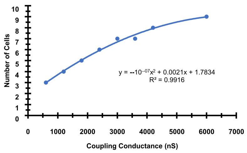
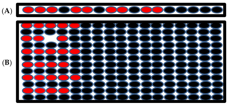
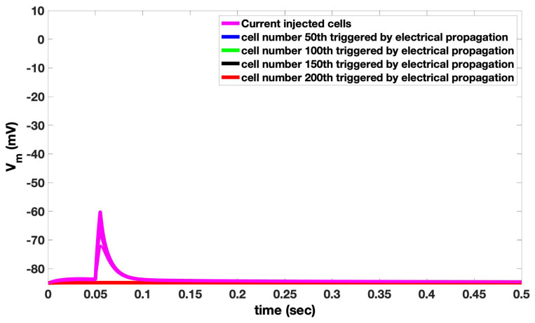
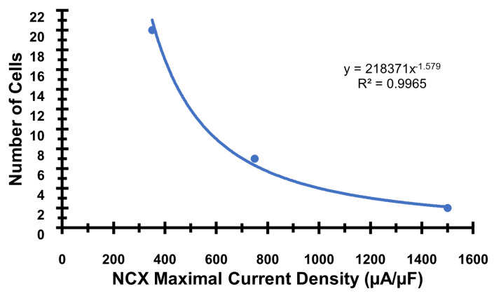
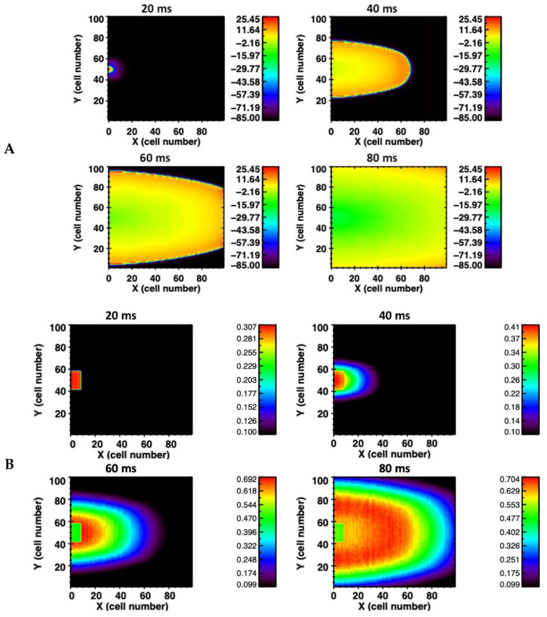
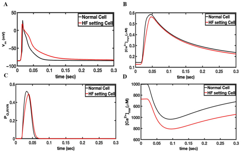
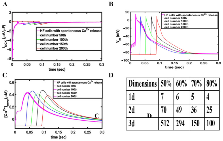
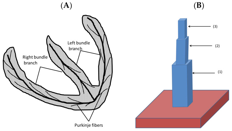
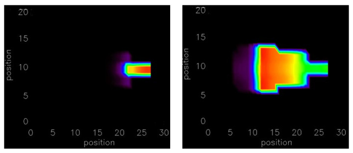
Cardiovascular disease is the leading cause of death worldwide due in a large part to arrhythmia. In order to understand how calcium dynamics play a role in arrhythmogenesis, normal and dysfunctional Ca signaling in a subcellular, cellular, and tissued level is examined using cardiac ventricular myocytes at a high temporal and spatial resolution using multiscale computational modeling. Ca sparks underlie normal excitation-contraction coupling. However, under pathological conditions, Ca sparks can combine to form Ca waves. These propagating elevations of (Ca) can activate an inward Na-Ca exchanger current (I) that contributes to early after-depolarization (EADs) and delayed after-depolarizations (DADs). However, how cellular currents lead to full depolarization of the myocardium and how they initiate extra systoles is still not fully understood. This study explores how many myocytes must be entrained to initiate arrhythmogenic depolarizations in biophysically detailed computational models. The model presented here suggests that only a small number of myocytes must activate in order to trigger an arrhythmogenic propagating action potential. These conditions were examined in 1-D, 2-D, and 3-D considering heart geometry. The depolarization of only a few hundred ventricular myocytes is required to trigger an ectopic depolarization. The number decreases under disease conditions such as heart failure. Furthermore, in geometrically restricted parts of the heart such as the thin muscle strands found in the trabeculae and papillary muscle, the number of cells needed to trigger a propagating depolarization falls even further to less than ten myocytes.
心血管疾病是全球范围内的主要死亡原因,在很大程度上归因于心律失常。为了了解钙动力学在心律失常发生中的作用,使用多尺度计算模型在亚细胞、细胞和组织水平上检查正常和功能障碍的 Ca 信号,具有高时间和空间分辨率。Ca 火花是正常兴奋-收缩偶联的基础。然而,在病理条件下,Ca 火花可以结合形成 Ca 波。这些传播的(Ca)升高可以激活内向的 Na-Ca 交换器电流(I),这有助于早期后除极(EAD)和延迟后除极(DAD)。然而,细胞电流如何导致心肌完全去极化以及它们如何引发额外的心动过速仍然不完全清楚。本研究探讨了在生物物理详细的计算模型中,需要多少心肌细胞被诱发以引发致心律失常的去极化。这里提出的模型表明,只需少量心肌细胞激活即可触发致心律失常的传播动作电位。这些条件在考虑心脏几何形状的 1-D、2-D 和 3-D 中进行了检查。只需去极化几百个心室肌细胞就足以引发异位去极化。在心力衰竭等疾病状态下,这个数量会减少。此外,在心脏的几何受限部分,如在小梁和乳头肌中发现的薄肌肉束,触发传播去极化所需的细胞数量甚至进一步减少到不到十个肌细胞。