• 文献检索
  • 文档翻译
  • 深度研究
  • 学术资讯
  • Suppr Zotero 插件Zotero 插件
  • 邀请有礼
  • 套餐&价格
  • 历史记录
应用&插件
Suppr Zotero 插件Zotero 插件浏览器插件Mac 客户端Windows 客户端微信小程序
定价
高级版会员购买积分包购买API积分包
服务
文献检索文档翻译深度研究API 文档MCP 服务
关于我们
关于 Suppr公司介绍联系我们用户协议隐私条款
关注我们

Suppr 超能文献

核心技术专利:CN118964589B侵权必究
粤ICP备2023148730 号-1Suppr @ 2026

文献检索

告别复杂PubMed语法,用中文像聊天一样搜索,搜遍4000万医学文献。AI智能推荐,让科研检索更轻松。

立即免费搜索

文件翻译

保留排版,准确专业,支持PDF/Word/PPT等文件格式,支持 12+语言互译。

免费翻译文档

深度研究

AI帮你快速写综述,25分钟生成高质量综述,智能提取关键信息,辅助科研写作。

立即免费体验

OCT 血管造影评估无临床糖尿病视网膜病变征象患者的黄斑病变:用于糖尿病视网膜病变早期筛查的放射组学特征。

Assessment of macular findings by OCT angiography in patients without clinical signs of diabetic retinopathy: radiomics features for early screening of diabetic retinopathy.

机构信息

Poostchi Ophthalmology Research Center, department of ophthalmology, Shiraz University of Medical Sciences, Shiraz, Iran.

Department of ophthalmology, Medical school, Shiraz University of Medical Sciences, Shiraz, Iran.

出版信息

BMC Ophthalmol. 2022 Jun 27;22(1):281. doi: 10.1186/s12886-022-02492-x.

DOI:10.1186/s12886-022-02492-x
PMID:35761260
原文链接:https://pmc.ncbi.nlm.nih.gov/articles/PMC9235114/
Abstract

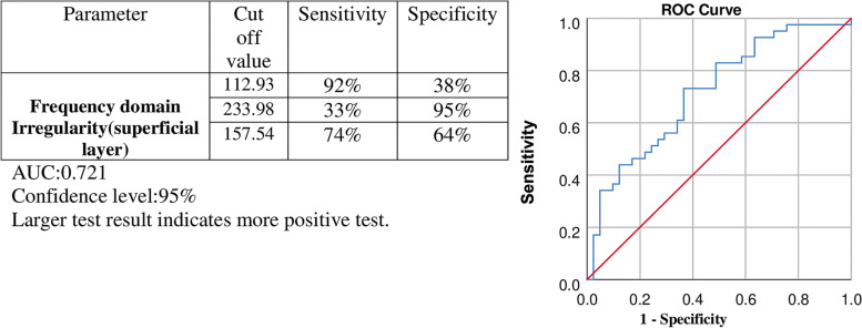
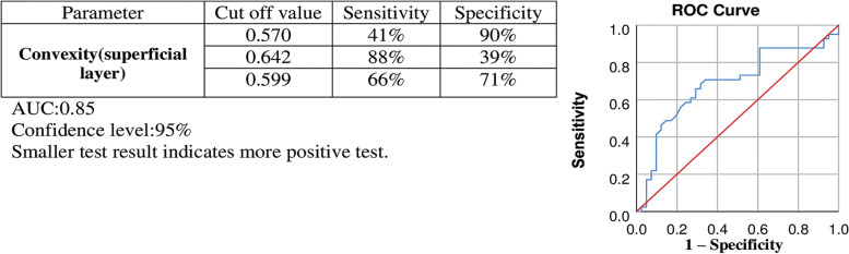
This cross-sectional study aimed to quantitatively analyze the optical coherence tomography angiography (OCTA) images using MATLAB-based software and evaluate the initial changes in macular vascular density and the distortion of the foveal avascular zone (FAZ), before the clinical appearance of diabetic retinopathy. For this purpose, 21 diabetic patients without any clinical features indicating DR, and 21 healthy individuals matched with patients based on their demographic characteristics were included. Macular thickness, macular vascular density, and morphological changes of FAZ were assessed using OCTA. The diagnostic ability of morphological parameters was evaluated by receiver operating curve analysis. The intraclass correlation coefficient (ICCC) index was used to check the consistency of the extracted values. There was no significant difference in age, gender, LogMAR visual acuity, spherical equivalent, and intra-ocular pressure amongst patients and controls. No correlation was found between age and the FAZ area as well as vascular density. The vascular structure of the superficial layer showed FAZ enlargement, reduced vascular density in the macular area, and significant deviations of FAZ shape parameters (convexity and Frequency Domain Irregularity) in patients compared with healthy individuals. Measurements were highly correlated between separate imaging sessions with ICCC of over 0.85 for all parameters. The represented data suggests that radiomics parameters can be applied as both an early screening tool and guidance for better follow-up of diabetic patients who have not had any sign of DR in fundoscopic exams.

摘要

本横断面研究旨在使用基于 MATLAB 的软件对光学相干断层扫描血管造影 (OCTA) 图像进行定量分析,并评估糖尿病视网膜病变 (DR) 临床症状出现之前黄斑血管密度的初始变化和中心凹无血管区 (FAZ) 的变形。为此,纳入了 21 名无任何 DR 临床特征的糖尿病患者和 21 名基于人口统计学特征与患者相匹配的健康个体。使用 OCTA 评估黄斑厚度、黄斑血管密度和 FAZ 的形态变化。通过接收者操作曲线分析评估形态学参数的诊断能力。采用组内相关系数 (ICCC) 指数检查提取值的一致性。患者和对照组在年龄、性别、LogMAR 视力、等效球镜和眼压方面无显著差异。年龄与 FAZ 面积和血管密度之间无相关性。与健康个体相比,浅层血管结构表现为 FAZ 扩大、黄斑区血管密度降低,以及 FAZ 形态参数 (凸度和频域不规则性) 显著偏离。所有参数的 ICCC 均超过 0.85,表明两次独立成像之间的测量高度相关。所代表的数据表明,放射组学参数可作为一种早期筛查工具,并为眼底检查尚无任何 DR 迹象的糖尿病患者提供更好的随访指导。

https://cdn.ncbi.nlm.nih.gov/pmc/blobs/4e6c/9235114/5076a091ccac/12886_2022_2492_Fig2_HTML.jpg
https://cdn.ncbi.nlm.nih.gov/pmc/blobs/4e6c/9235114/91f0b7bae148/12886_2022_2492_Fig1_HTML.jpg
https://cdn.ncbi.nlm.nih.gov/pmc/blobs/4e6c/9235114/5076a091ccac/12886_2022_2492_Fig2_HTML.jpg
https://cdn.ncbi.nlm.nih.gov/pmc/blobs/4e6c/9235114/91f0b7bae148/12886_2022_2492_Fig1_HTML.jpg
https://cdn.ncbi.nlm.nih.gov/pmc/blobs/4e6c/9235114/5076a091ccac/12886_2022_2492_Fig2_HTML.jpg

相似文献

1
Assessment of macular findings by OCT angiography in patients without clinical signs of diabetic retinopathy: radiomics features for early screening of diabetic retinopathy.OCT 血管造影评估无临床糖尿病视网膜病变征象患者的黄斑病变:用于糖尿病视网膜病变早期筛查的放射组学特征。
BMC Ophthalmol. 2022 Jun 27;22(1):281. doi: 10.1186/s12886-022-02492-x.
2
Vessel Density around Foveal Avascular Zone as a Potential Imaging Biomarker for Detecting Preclinical Diabetic Retinopathy: An Optical Coherence Tomography Angiography Study.黄斑中心无血管区周围血管密度作为一种潜在的影像学生物标志物用于检测临床前期糖尿病视网膜病变: 一项光学相干断层扫描血管造影研究。
Semin Ophthalmol. 2020 Aug 17;35(5-6):316-323. doi: 10.1080/08820538.2020.1845386. Epub 2020 Dec 1.
3
Evaluation of Automatically Quantified Foveal Avascular Zone Metrics for Diagnosis of Diabetic Retinopathy Using Optical Coherence Tomography Angiography.基于光学相干断层扫描血管造影的自动量化中心凹无血管区指标对糖尿病视网膜病变的诊断评估。
Invest Ophthalmol Vis Sci. 2018 May 1;59(6):2212-2221. doi: 10.1167/iovs.17-23498.
4
Macular vessels density in diabetic retinopathy: quantitative assessment using optical coherence tomography angiography.糖尿病视网膜病变中的黄斑血管密度:使用光学相干断层扫描血管造影术进行定量评估。
Int Ophthalmol. 2019 Aug;39(8):1845-1859. doi: 10.1007/s10792-018-1013-0. Epub 2018 Sep 7.
5
Optical coherence tomography angiography analysis of foveal microvascular changes and inner retinal layer thinning in patients with diabetes.糖尿病患者黄斑区微血管改变及内层视网膜变薄的光相干断层扫描血管造影分析。
Br J Ophthalmol. 2018 Sep;102(9):1226-1231. doi: 10.1136/bjophthalmol-2017-311149. Epub 2017 Dec 19.
6
Linking Retinal Microvasculature Features With Severity of Diabetic Retinopathy Using Optical Coherence Tomography Angiography.使用光学相干断层扫描血管造影术将视网膜微血管特征与糖尿病性视网膜病变的严重程度相联系。
Invest Ophthalmol Vis Sci. 2016 Jul 1;57(9):OCT519-25. doi: 10.1167/iovs.15-18901.
7
Correlation Analysis between Foveal Avascular Zone and Peripheral Ischemic Index in Diabetic Retinopathy: A Pilot Study.糖尿病视网膜病变中黄斑无血管区与周边缺血指数的相关性分析:一项初步研究
Ophthalmol Retina. 2018 Jan;2(1):46-52. doi: 10.1016/j.oret.2017.05.007. Epub 2017 Jul 26.
8
Early microvascular retinal changes in optical coherence tomography angiography in patients with type 1 diabetes mellitus.1型糖尿病患者光学相干断层扫描血管造影中的早期视网膜微血管变化。
Acta Ophthalmol. 2017 Dec;95(8):e751-e755. doi: 10.1111/aos.13404. Epub 2017 Feb 16.
9
Retinal neurodegeneration, macular circulation and morphology of the foveal avascular zone in diabetic patients: quantitative cross-sectional study using OCT-A.糖尿病患者的视网膜神经退行性变、黄斑循环和中心无血管区形态:使用 OCT-A 的定量横断面研究。
Acta Ophthalmol. 2021 Nov;99(7):e1135-e1140. doi: 10.1111/aos.14754. Epub 2021 Jan 10.
10
Quantification of Diabetic Macular Ischemia Using Optical Coherence Tomography Angiography and Its Relationship with Visual Acuity.利用光相干断层扫描血管造影术对糖尿病性黄斑水肿进行定量分析及其与视力的关系。
Ophthalmology. 2017 Feb;124(2):235-244. doi: 10.1016/j.ophtha.2016.10.008. Epub 2016 Nov 23.

引用本文的文献

1
Recent advances and applications of optical coherence tomography angiography in diabetic retinopathy.光学相干断层扫描血管造影在糖尿病视网膜病变中的最新进展与应用
Front Endocrinol (Lausanne). 2025 Apr 16;16:1438739. doi: 10.3389/fendo.2025.1438739. eCollection 2025.
2
Radiomics-Based OCT Analysis of Choroid Reveals Biomarkers of Central Serous Chorioretinopathy.基于影像组学的脉络膜光学相干断层扫描分析揭示了中心性浆液性脉络膜视网膜病变的生物标志物。
Transl Vis Sci Technol. 2025 Apr 1;14(4):23. doi: 10.1167/tvst.14.4.23.
3
Enhancing Retinal Resilience: The Neuroprotective Promise of BDNF in Diabetic Retinopathy.

本文引用的文献

1
An Open-source Image Analysis Toolbox for Quantitative Retinal Optical Coherence Tomography Angiography.用于定量视网膜光学相干断层扫描血管造影的开源图像分析工具箱。
J Biomed Phys Eng. 2024 Feb 1;14(1):31-42. doi: 10.31661/jbpe.v0i0.2106-1349. eCollection 2024 Feb.
2
Diabetic Retinopathy and BDNF: A Review on Its Molecular Basis and Clinical Applications.糖尿病视网膜病变与脑源性神经营养因子:关于其分子基础及临床应用的综述
J Ophthalmol. 2020 May 18;2020:1602739. doi: 10.1155/2020/1602739. eCollection 2020.
3
OCT Angiography Biomarkers for Predicting Visual Outcomes after Ranibizumab Treatment for Diabetic Macular Edema.
增强视网膜韧性:脑源性神经营养因子在糖尿病视网膜病变中的神经保护前景。
Life (Basel). 2025 Feb 9;15(2):263. doi: 10.3390/life15020263.
4
Sub-Clinical Regional Macular Changes in Self-Reported Diabetic Subjects: Vascular and Retinal Layer Analysis Using Swept-Source OCT.自我报告的糖尿病患者的亚临床区域黄斑变化:使用扫频光学相干断层扫描进行血管和视网膜层分析
Diabetes Metab Syndr Obes. 2025 Feb 6;18:363-372. doi: 10.2147/DMSO.S504882. eCollection 2025.
5
Radiomics in ophthalmology: a systematic review.眼科中的放射组学:一项系统综述。
Eur Radiol. 2025 Jan;35(1):542-557. doi: 10.1007/s00330-024-10911-4. Epub 2024 Jul 21.
6
Radiomics-Based Prediction of Anti-VEGF Treatment Response in Neovascular Age-Related Macular Degeneration With Pigment Epithelial Detachment.基于影像组学对伴有色素上皮脱离的新生血管性年龄相关性黄斑变性抗VEGF治疗反应的预测
Transl Vis Sci Technol. 2023 Oct 3;12(10):3. doi: 10.1167/tvst.12.10.3.
7
Artificial Intelligence-Assisted Processing of Anterior Segment OCT Images in the Diagnosis of Vitreoretinal Lymphoma.人工智能辅助处理眼前节光学相干断层扫描(OCT)图像在玻璃体视网膜淋巴瘤诊断中的应用
Diagnostics (Basel). 2023 Jul 23;13(14):2451. doi: 10.3390/diagnostics13142451.
8
Essential Role of Multi-Omics Approaches in the Study of Retinal Vascular Diseases.多组学方法在视网膜血管疾病研究中的重要作用。
Cells. 2022 Dec 26;12(1):103. doi: 10.3390/cells12010103.
9
Volume-based, layer-independent, disease-agnostic detection of abnormal retinal reflectivity, nonperfusion, and neovascularization using structural and angiographic OCT.使用结构和血管造影光学相干断层扫描(OCT)基于体积、不依赖层、与疾病无关地检测视网膜反射率异常、无灌注和新生血管形成。
Biomed Opt Express. 2022 Aug 22;13(9):4889-4906. doi: 10.1364/BOE.469308. eCollection 2022 Sep 1.
预测雷珠单抗治疗糖尿病性黄斑水肿后视力预后的光学相干断层扫描血管造影生物标志物
Ophthalmol Retina. 2019 Oct;3(10):826-834. doi: 10.1016/j.oret.2019.04.027. Epub 2019 May 7.
4
Methods.方法。
Can J Diabetes. 2018 Apr;42 Suppl 1:S6-S9. doi: 10.1016/j.jcjd.2017.10.002.
5
A basis for customising perimetric locations within the macula in glaucoma.青光眼黄斑区内视野检查位置定制的基础。
Ophthalmic Physiol Opt. 2018 Mar;38(2):164-173. doi: 10.1111/opo.12435. Epub 2018 Jan 8.
6
Radiomics: the bridge between medical imaging and personalized medicine.放射组学:医学影像与个性化医疗之间的桥梁。
Nat Rev Clin Oncol. 2017 Dec;14(12):749-762. doi: 10.1038/nrclinonc.2017.141. Epub 2017 Oct 4.
7
Retinal Microvasculature in Nonproliferative Diabetic Retinopathy: Automated Quantitative Optical Coherence Tomography Angiography Assessment.非增殖性糖尿病视网膜病变中的视网膜微血管:自动定量光学相干断层扫描血管造影评估
Ophthalmic Res. 2017;58(3):131-141. doi: 10.1159/000471885. Epub 2017 May 25.
8
Optical coherence tomography angiography analysis of retinal vascular plexuses and choriocapillaris in patients with type 1 diabetes without diabetic retinopathy.1型糖尿病无糖尿病视网膜病变患者视网膜血管丛和脉络膜毛细血管的光学相干断层扫描血管造影分析
Acta Diabetol. 2017 Jul;54(7):695-702. doi: 10.1007/s00592-017-0996-8. Epub 2017 May 5.
9
Diabetic Macular Edema Pathophysiology: Vasogenic versus Inflammatory.糖尿病性黄斑水肿的病理生理学:血管源性与炎症性
J Diabetes Res. 2016;2016:2156273. doi: 10.1155/2016/2156273. Epub 2016 Sep 28.
10
Global Estimates on the Number of People Blind or Visually Impaired by Diabetic Retinopathy: A Meta-analysis From 1990 to 2010.全球因糖尿病视网膜病变致盲或视力受损人数的估计:1990 年至 2010 年的荟萃分析。
Diabetes Care. 2016 Sep;39(9):1643-9. doi: 10.2337/dc15-2171.